Section 06 of 10
Structures as 3D objects
Let's apply this depth framework to common dermoscopic structures.
Pigment network
What it is: A honeycomb pattern of pigmented lines.
3D interpretation: You're seeing the pigmented basal layer of the epidermis, which drapes over dermal papillae. The "holes" in the network are the tips of the papillae; the "lines" are the rete ridges surrounding them.
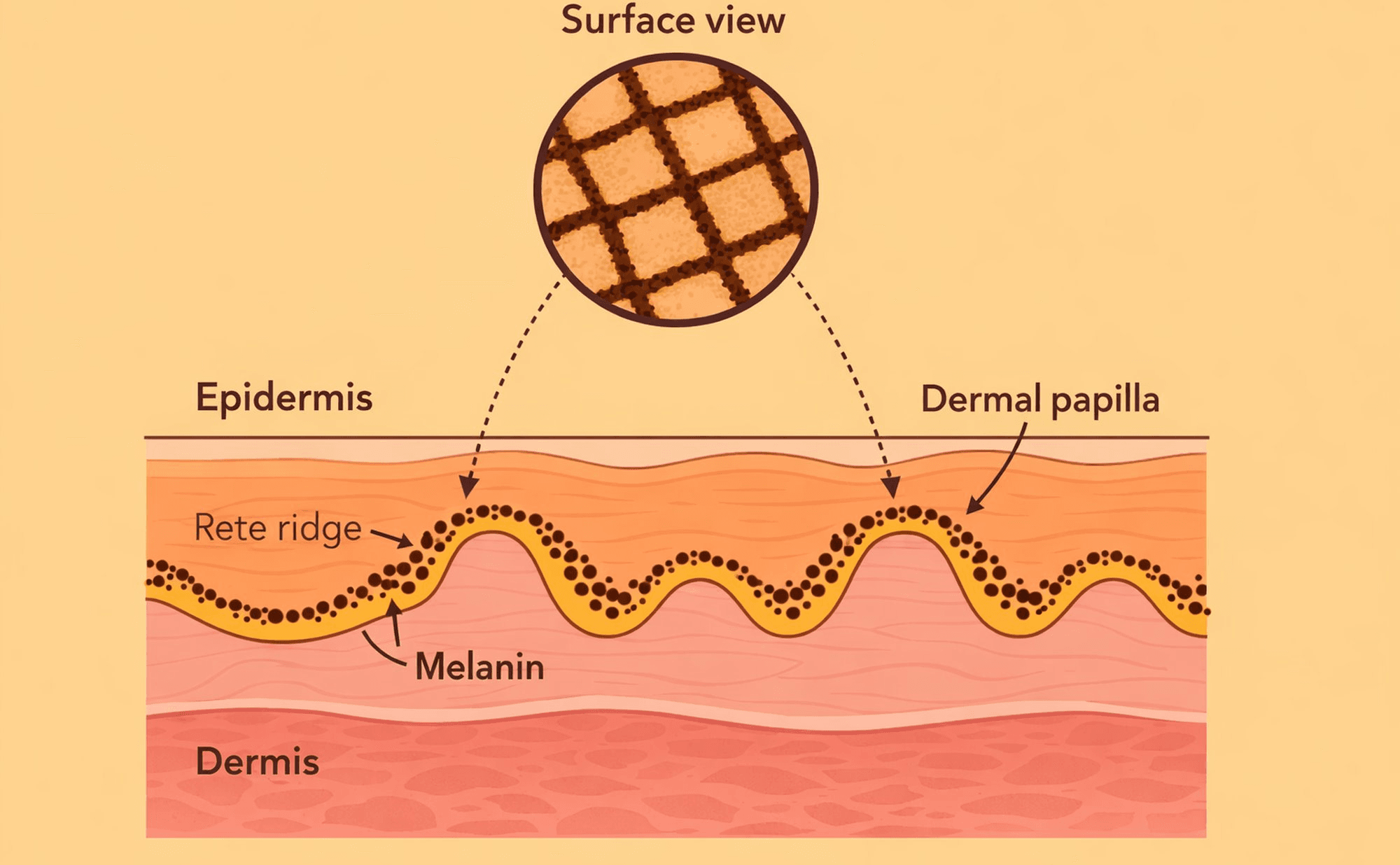
Rete ridge architecture — the 3D structure that creates the pigment network pattern seen in dermoscopy.
Depth cue: True pigment network sits at the dermo-epidermal junction. It should have an "embedded" appearance, not a surface appearance.
Dots and globules
What they are: Small round or oval pigmented structures.
3D interpretation: Dots are small aggregates of melanin or melanocytes. Globules are larger aggregates, often representing nests of cells.
Depth cue: Their colour indicates their level. Dark dots are higher; grey-brown globules are deeper.
Blue-grey areas
What they are: Areas with a steel-blue or grey hue.
3D interpretation: Pigment deep in the dermis. Light scattering through overlying tissue shifts the apparent colour toward blue — this is known as the Tyndall effect, similar to why veins appear blue through skin.
Blue-grey colour always indicates dermal depth. This is one of the most reliable depth cues in dermoscopy and has important implications for lesion assessment.
White structures
What they are: Bright white lines, areas, or "shiny" structures.
3D interpretation: These often represent fibrosis, regression, or specific tissue changes that reflect light differently. "Shiny white structures" (visible only in polarised light) indicate stromal changes at depth.
Depth cue: White structures usually indicate altered tissue architecture, often with clinical significance.
Which of the following statements about pigment network are correct? Select all that apply.