Section 04 of 10
The mental shift: from pattern to structure
Beginners often describe dermoscopic features in two-dimensional terms: "I see a brown blob," "There's a network pattern," "The edges look irregular."
Experts describe the same features in three-dimensional terms: "There's pigment aggregated at the dermo-epidermal junction," "The network reflects rete ridge architecture," "The asymmetry suggests uneven growth in multiple planes."
The information entering the eye is identical. The interpretation differs because the expert has learned to decode spatial relationships.
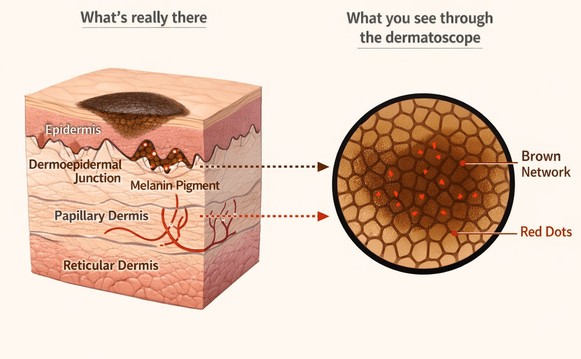
What's really there vs what you see — how 3D skin structures map to the 2D dermoscopic image.
A simple exercise
Look at a dermoscopic image and ask yourself three questions, working from top to bottom:
- What sits on the surface? Scale, crust, surface texture
- What sits within the epidermis? Pigmented network, dots, globules
- What sits deeper? Blue-grey areas, vessels, white structures
This simple layering exercise begins to train your brain to see depth rather than flat pattern.