Section 03 of 10
How the dermatoscope creates its view
Understanding the instrument helps you interpret its output.
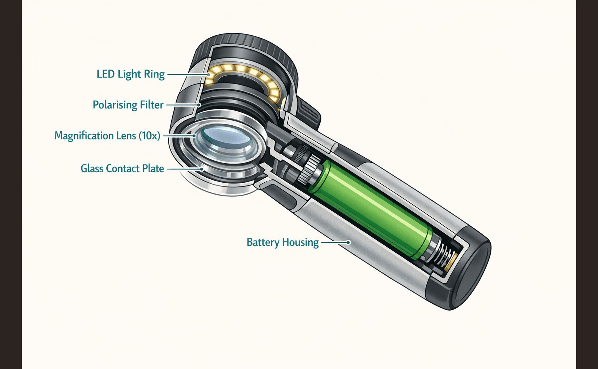
Internal components of a typical polarised dermatoscope.
Real dermatoscopes vary in shape and size between manufacturers. The image you see through the lens will also differ depending on whether you use polarised or non-polarised (contact) mode — both are covered later in this section.
Magnification
Standard dermatoscopes provide 10× magnification. This makes features visible that are invisible to the naked eye, but it also changes your spatial reference. A structure that appears 2mm across in the dermatoscope is actually 0.2mm in reality.
Train yourself to think in dermoscopic scale. "Small" and "large" take on different meanings at 10× magnification.
Light penetration
Light enters the skin and interacts with structures at different depths:
- Surface structures reflect light directly — they appear bright and well-defined
- Mid-depth structures scatter light — they appear softer, with gradients
- Deep structures absorb light selectively — they appear as colour shifts (blue-grey tones)
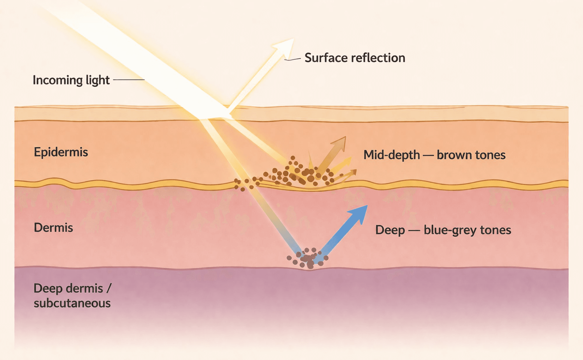
Light penetration at three depths — surface (bright, crisp), mid-depth (softer, scattered), and deep (blue-grey colour shift).
The colour–depth relationship shown here is a useful guide, not a precise rule. In practice, lesion type, thickness, and your dermoscopic technique all influence the colours you see.
Colour is a depth cue. Brown at the surface looks different from brown at the junction, which looks different from pigment in the dermis. Learning to read these differences is at the heart of dermoscopy.
Polarised vs non-polarised
- Non-polarised (contact) dermoscopy requires gel or fluid and shows surface features well
- Polarised (non-contact) dermoscopy eliminates surface reflection and reveals deeper structures
Many dermatoscopes offer both modes. Learning to interpret each takes practice.
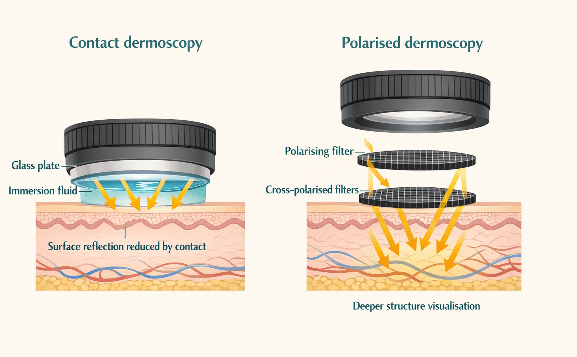
Contact vs polarised dermoscopy — two approaches to eliminating surface reflection and revealing subsurface structures.
Both modes can reveal overlapping features. Polarised dermoscopy does not literally see deeper layers — it reduces surface glare, making structures visible that contact dermoscopy may obscure.
A structure appears bright, well-defined, and crisp through the dermatoscope. What does this suggest about where it sits?