TADA Simulator
Practice the three-step algorithm with training scenarios. Work through each case at your own pace — every answer includes educational feedback to reinforce your learning.
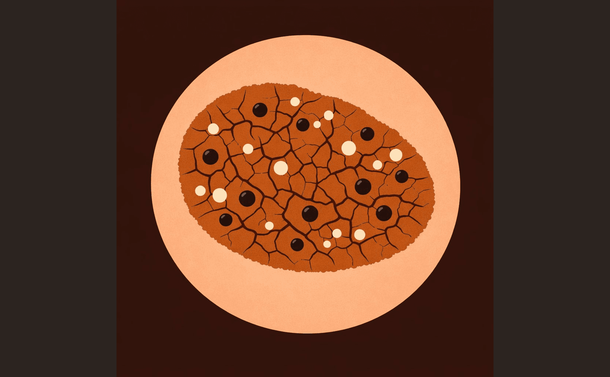
72-year-old patient. Lesion on the trunk, present for several years, slowly enlarging. Not painful or itchy.
A well-defined, slightly raised lesion with a waxy surface. Under dermoscopy, you observe multiple small round white inclusions and darker round openings across the surface. The overall pattern has a textured, "stuck-on" quality with a brain-like arrangement of ridges and fissures.
Benign pattern recognition
In this training scenario, does the lesion show unequivocal features of…
How the simulator works
Pattern recognition
Each scenario begins with Step 1: does the lesion show unequivocal features of seborrhoeic keratosis, cherry angioma, or dermatofibroma?
Organisation assessment
If no benign pattern is recognised, Step 2 asks whether the lesion appears organised (symmetric, uniform) or disorganised (asymmetric, chaotic).
Feature recognition
Step 3 checks for seven high-risk features. Any single feature present is educationally significant — you do not need to find multiple features.
All 8 scenarios are educational only. Click any image to examine it in detail. Each dermoscopic illustration is designed to clearly demonstrate the features described in the scenario, so you can practise identifying them before encountering them in clinical practice.