Section 03 of 09
Benign structures you will see everywhere
Beyond the pigment network, there are several other structures that appear frequently in healthy skin and benign lesions. Learning to recognise them quickly will save you time and build your confidence.
Comedo-like openings
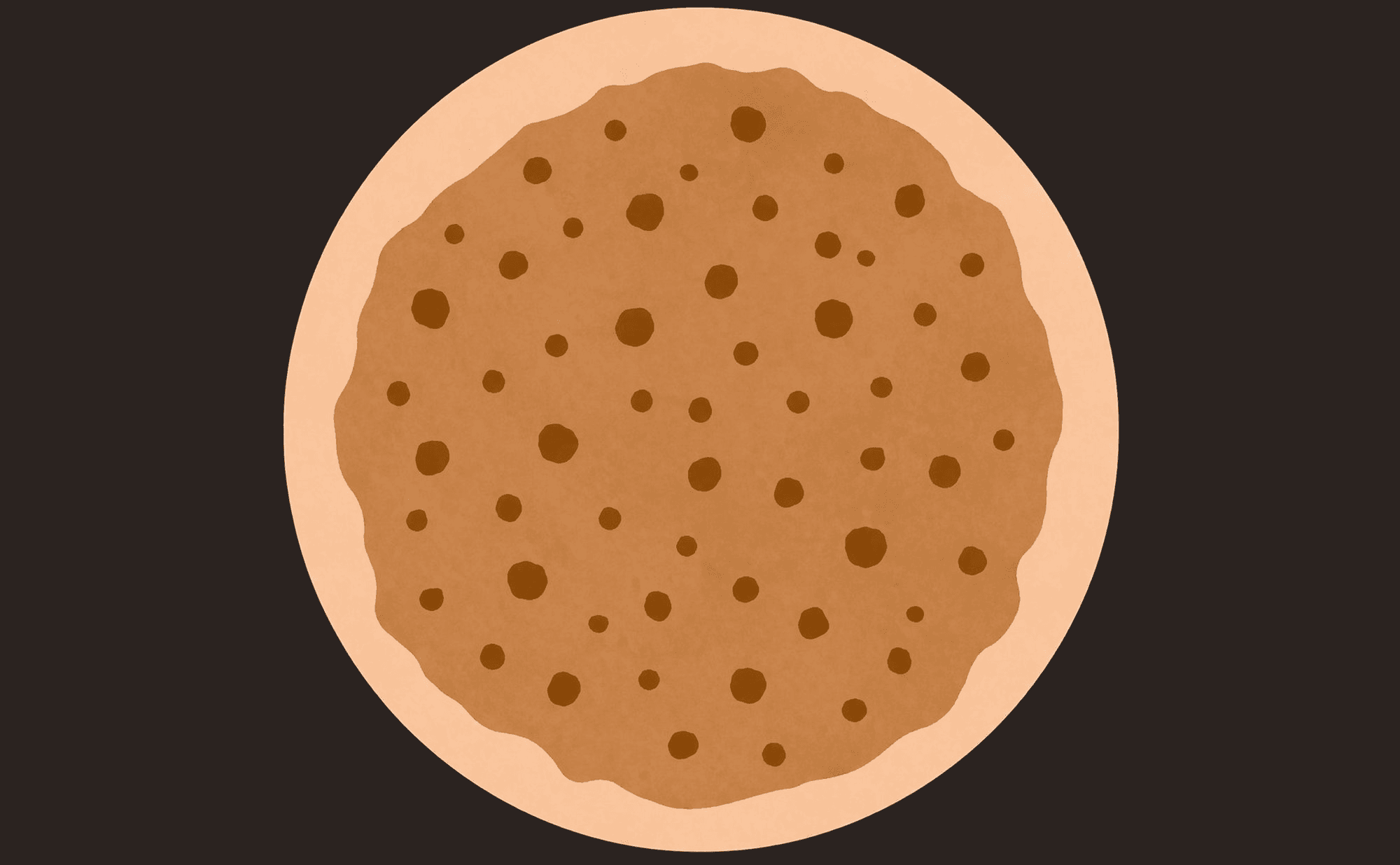
Comedo-like openings: dark yellowish-brown keratin plugs within a pigmented lesion
These look like small, round or oval yellowish-brown plugs embedded in the surface of a lesion — rather like blackheads. The name is deliberate: they resemble comedones (blackheads), though they are actually keratin-filled pits in the skin surface.
You will encounter comedo-like openings most often in seborrhoeic keratosis — the extremely common benign growths covered in depth in Module 4. For now, simply learn to recognise the appearance: round, well-defined yellowish-brown holes that give the surface a pitted or cratered look.
Milia-like cysts
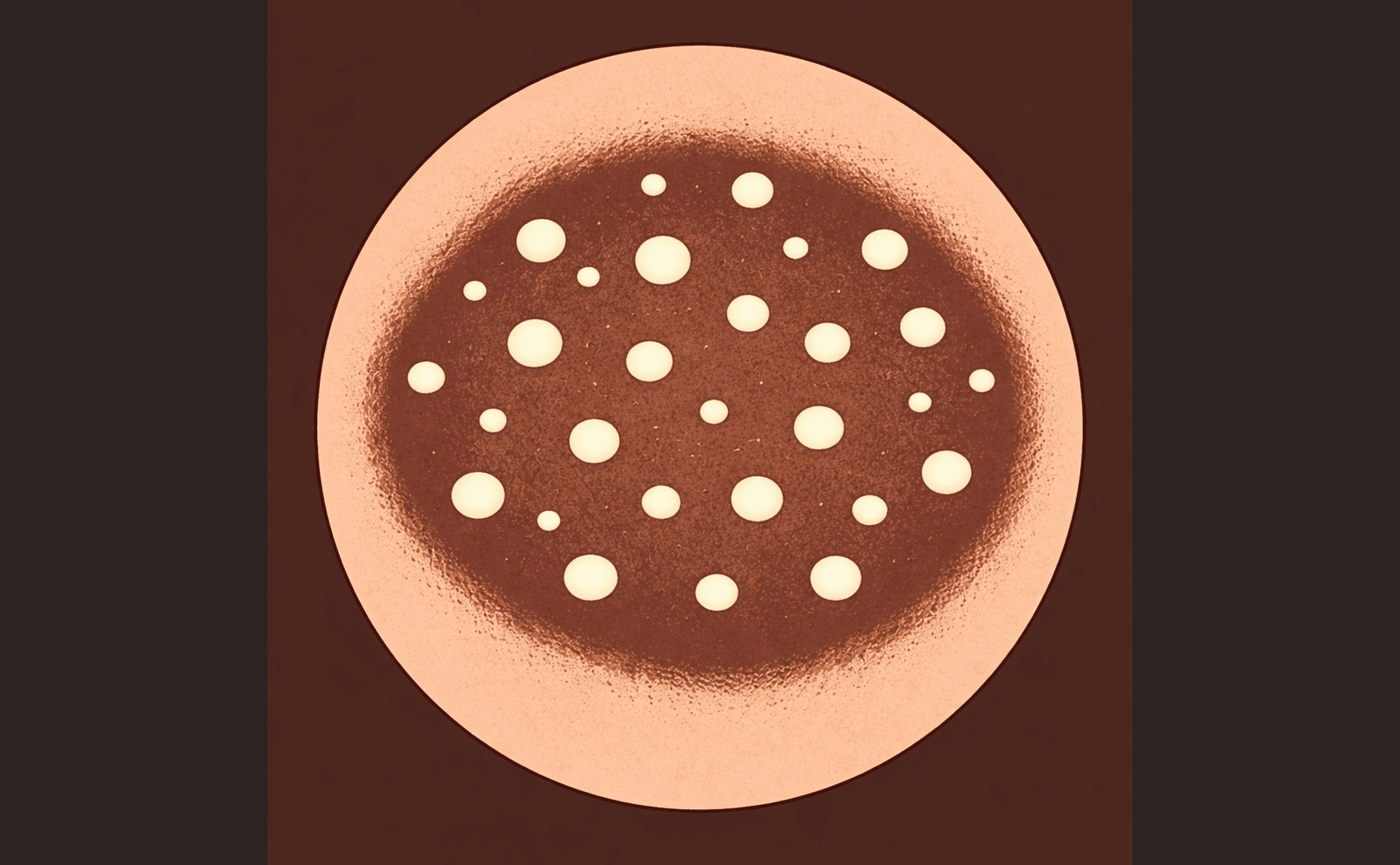
Milia-like cysts: bright white-yellow keratin pearls on a pigmented surface
These are small, bright white or yellowish-white round dots — like tiny pearls scattered across the surface of a lesion. They are caused by small keratin cysts trapped within the skin.
Like comedo-like openings, milia-like cysts are a hallmark feature of seborrhoeic keratosis. Seeing both together — the brown plugs and the white pearls — is one of the most reliable benign pattern combinations in dermoscopy.
Fissures and ridges
Some benign lesions have a surface texture that looks like the folds of a brain — a pattern of winding ridges separated by narrow grooves. This cerebriform pattern (brain-like pattern) is another classic sign of seborrhoeic keratosis.
Sharp demarcation
Many benign lesions have clean, well-defined borders. The edge of the lesion is crisp and distinct — you can easily see where the lesion ends and normal skin begins. While sharp demarcation alone does not confirm a benign diagnosis, it is a feature that contributes to the overall impression of an organised, well-behaved lesion.
Comedo-like openings (brown plugs) and milia-like cysts (white pearls) appearing together are one of the most reliable benign pattern combinations. When you see both, seborrhoeic keratosis is the most likely explanation — though you should still assess the full picture.
Red lacunae
In vascular lesions, you may see well-defined round or oval spaces filled with red, dark red, or purple colour. These lacunae are dilated blood-filled spaces, and they are the hallmark of cherry angioma — small red benign growths that are covered in Module 5.
Which combination of structures is considered one of the most reliable indicators of a benign seborrhoeic keratosis?