Section 03 of 08
Classic dermoscopic features
The dermoscopic pattern of a dermatofibroma reflects its layered structure: a dense fibrous core in the centre, with reactive changes in the overlying and surrounding epidermis.
Central white scar-like patch
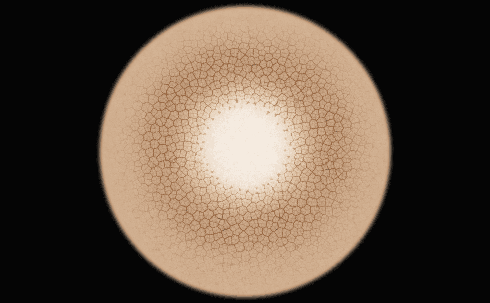
The hallmark feature is a central white patch — a well-defined area of white, structureless dermoscopic appearance occupying the centre of the lesion. This white zone corresponds to the dense collagen and fibrosis of the dermal nodule pressing upward against the overlying epidermis.
The white patch is not simply pale skin. Under polarised dermoscopy, it can appear quite bright — almost glistening — because the dense collagen fibres scatter polarised light in a distinctive way. This is an example of how fibrosis creates visible white structures under the dermatoscope, a principle that applies to other lesion types as well.
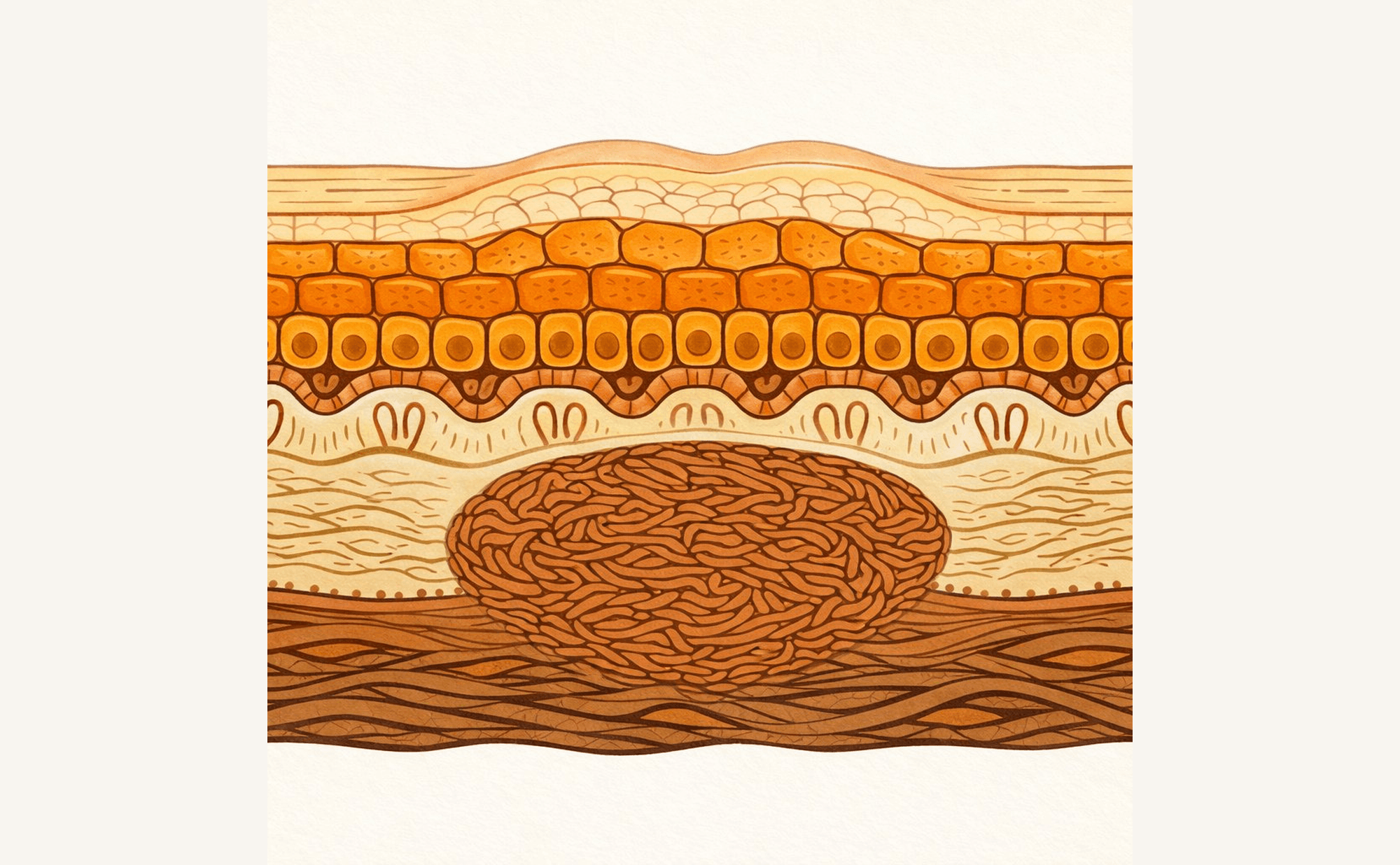
Why the white patch appears: the dense fibrous core pushes upward against the epidermis, creating the structureless white zone seen under dermoscopy
Peripheral pigment network
Surrounding the central white area, most dermatofibromas display a delicate pigment network. This network is typically fine and regular, with thin brown lines forming a mesh pattern that fades gradually toward the edges of the lesion.
The pigment network in a dermatofibroma is not caused by melanocyte proliferation — it is a reactive change. The fibrous core underneath pushes the epidermis upward, elongating the rete ridges and concentrating the melanin that is naturally present. This creates the network pattern as a secondary effect of the underlying fibrosis.
The network is usually most visible at the periphery, forming a ring or halo around the central white zone. It often has a delicate, almost fingerprint-like quality — finer than the bold network you might see in a melanocytic naevus.
The classic combination
Classic dermatofibroma: central white patch with peripheral pigment network — the bullseye pattern
The combination of these two features — central white patch surrounded by peripheral pigment network — is highly characteristic. It creates a target-like or bullseye pattern: white centre, brown ring, fading edges.
What causes the central white patch in a dermatofibroma?