Section 03 of 08
Classic dermoscopic features
The dermoscopic appearance of a cherry angioma is dominated by its vascular architecture. You are looking directly at dilated blood-filled spaces through the magnified lens.
Lacunae — the hallmark feature
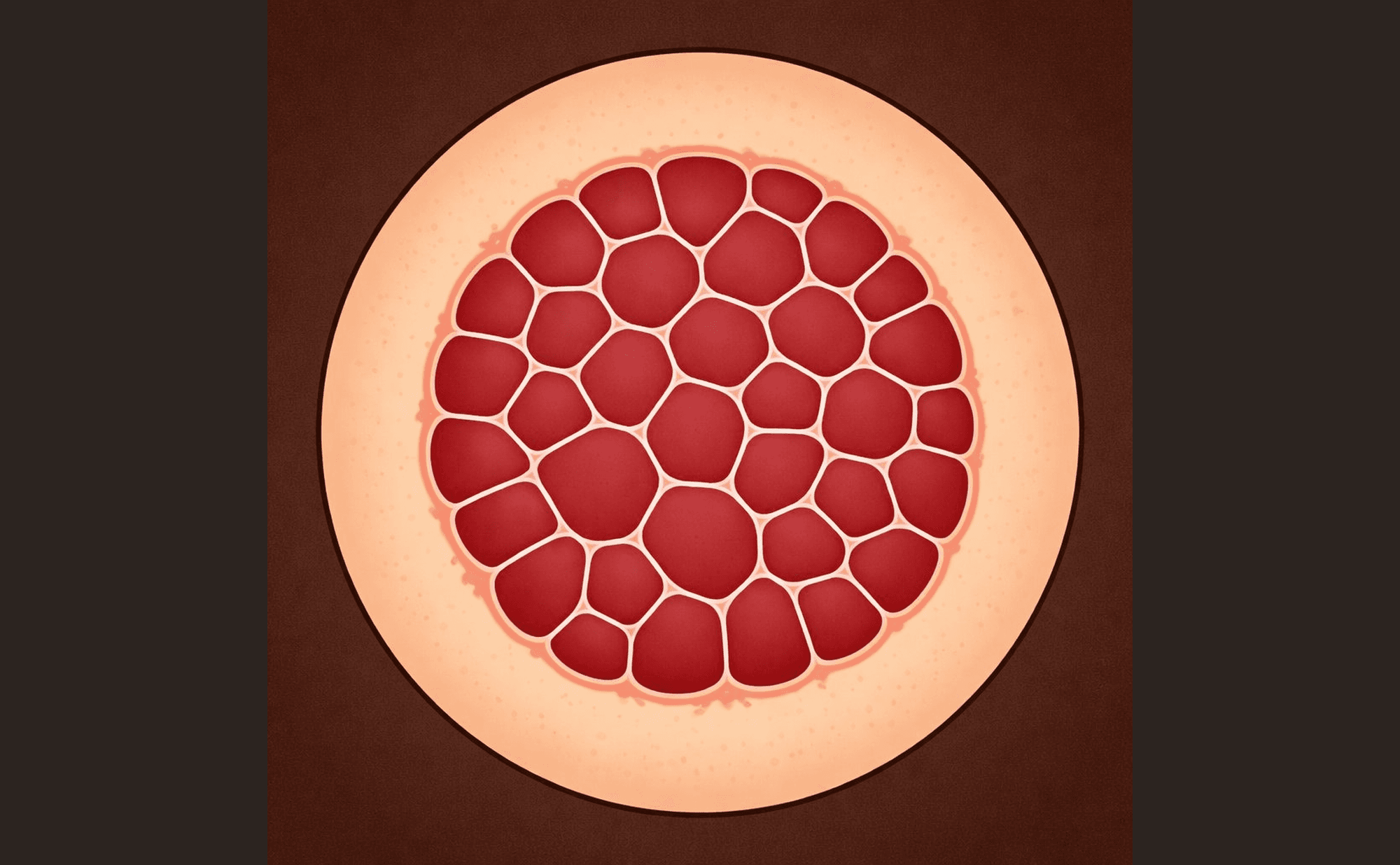
Classic cherry angioma: red-purple lacunae with pale septa — the 'stained glass' pattern
The defining feature of a cherry angioma is the presence of lacunae — round to oval compartments filled with red or red-purple colour. These correspond to dilated vascular spaces packed with red blood cells. Think of them as small pools of blood separated by thin walls.
Lacunae can vary in size within the same lesion. Some are large enough to see individually as distinct round or oval shapes. Others are smaller and may merge together. The colour within each lacuna is usually uniform — a rich red or deep red-purple depending on how much blood is present and how deep the vessels sit.
Pale septa
Between the lacunae, you will often see thin pale lines — these are the septa, the connective tissue walls that separate the blood-filled compartments. They create a mesh-like or honeycomb appearance when viewed from above. The septa appear pale or white because they are made of fibrous tissue rather than blood.
Not all cherry angiomas show clearly visible septa. In smaller lesions, the vascular spaces may be too tightly packed to see individual dividing walls. But when they are visible, septa confirm the lacunar architecture and strengthen your identification.
Homogeneous red pattern
Smaller or early cherry angiomas may appear as a uniform red area without visible internal structure. This homogeneous pattern occurs when the vascular spaces are too small to resolve as individual lacunae — the whole lesion simply looks like a well-defined red dot.
This is still consistent with a cherry angioma, provided the colour is uniform and the borders are sharp. As the lesion grows, lacunae often become visible over time.
Sharp demarcation
Cherry angiomas have a crisp, well-defined edge. The transition from the red of the lesion to the normal surrounding skin is abrupt — there is no gradual fading, no blurring, no irregular extension into the surrounding tissue. This sharp boundary reflects the contained nature of the vascular proliferation.
Which feature is most characteristic of a cherry angioma under dermoscopy?