How to use this glossary
This glossary defines every dermoscopic term used across the BorelliScopie® platform. Each entry provides a plain-language description, what you see through the dermatoscope, an illustrated example, and the lesions where the feature is most commonly encountered.
Browse alphabetically, or use it as a quick lookup when you encounter an unfamiliar term in the learning modules.
Each entry follows the BorelliScopie three-layer approach: plain description first, then what you see, then the clinical associations. The formal dermoscopic term is the heading — but understanding comes from the description beneath it.
A
Annular-granular pattern
Plain language: Grey dots arranged in rings around hair follicles on the face.
What you see: Fine grey granules forming circular patterns around follicular openings — like tiny necklaces encircling each follicle. Found on sun-damaged facial skin.
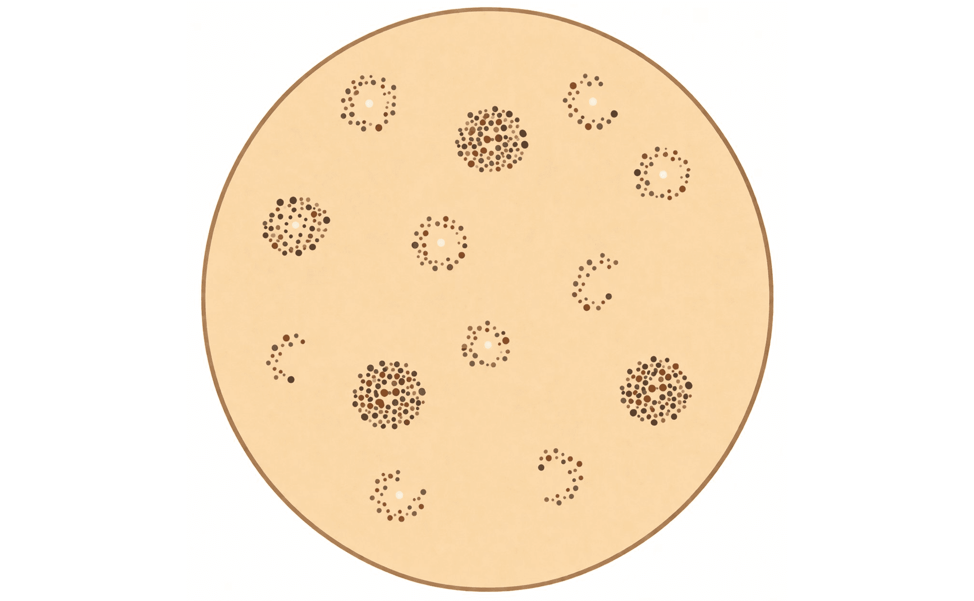
Annular-granular pattern: grey granules forming ring-like arrangements around follicular openings — characteristic of lentigo maligna on facial skin
Associated with: Lentigo maligna (characteristic feature). See Blue, Black & Grey and Malignant Patterns.
Arborising vessels
Plain language: Bright red blood vessels that branch like tree limbs.
What you see: Sharply focused, bright red vessels with hierarchical branching — a larger trunk divides into progressively finer branches. Crisp and well-defined against a pale background.
What you see
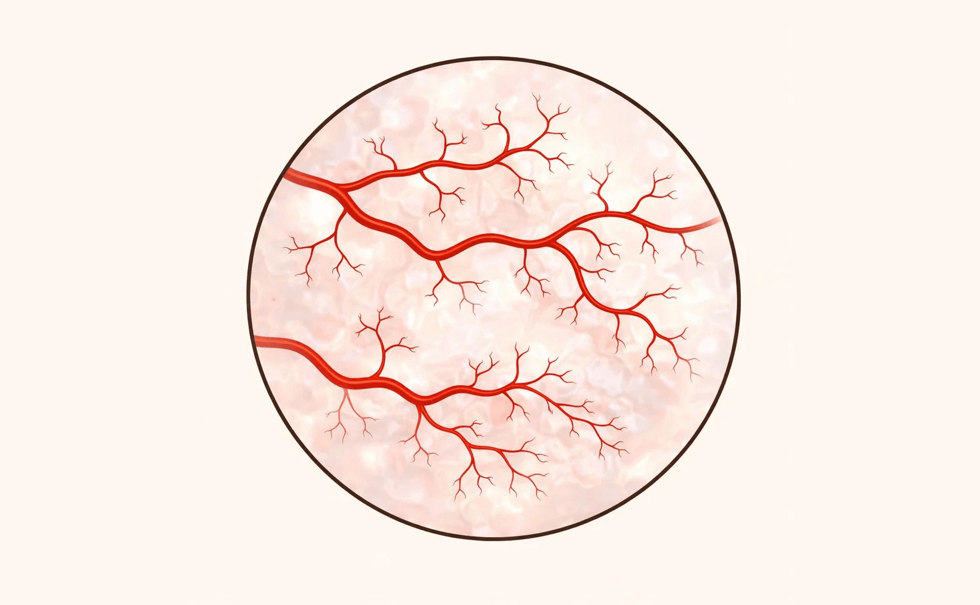
Arborising vessels: tortuous bright red vessels with dichotomous branching and progressive calibre reduction, characteristic of basal cell carcinoma
What it resembles

A bare winter tree — trunk dividing dichotomously into finer branches, the living analogy for arborising vessels
Associated with: Basal cell carcinoma (highly characteristic). See Vessels and Non-Melanoma Skin Cancer.
Asymmetry
Plain language: When one half of a lesion does not match the other.
What you see: Differences in colour, structure, or pattern when comparing opposite sides of a lesion. Assessed in two perpendicular axes. Asymmetry of structure (not just shape) is the most diagnostically significant form.
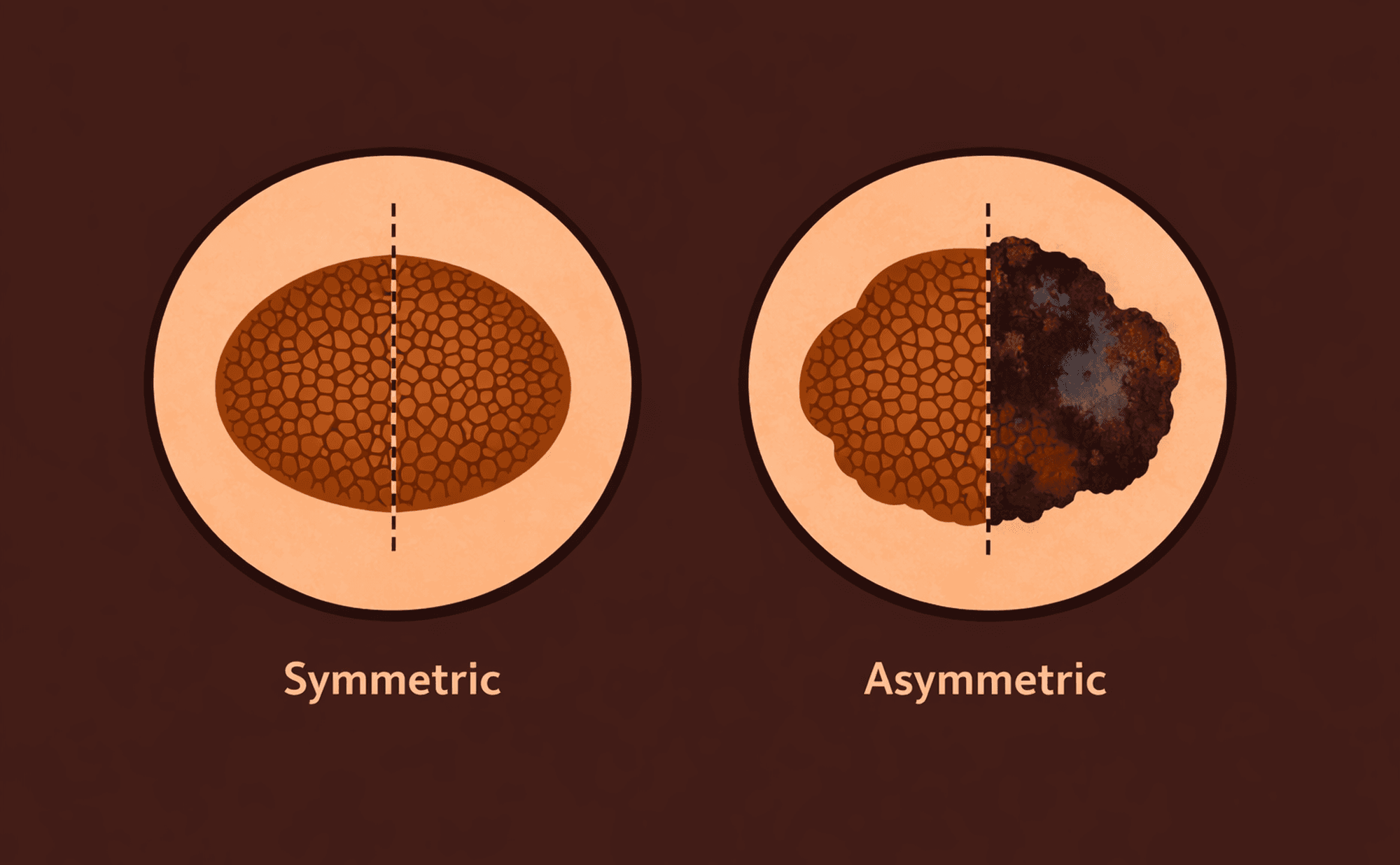
Left: symmetric lesion — similar structures in both halves. Right: asymmetric lesion — different colours, structures, and patterns when halved
Associated with: Melanoma, atypical naevi. See Malignant Patterns.
Atypical network
Plain language: Irregular honeycomb pattern with uneven lines and spaces.
What you see: Pigment network with variable line thickness, irregular mesh size, broken or branched lines, and focal thickening. The rhythm and regularity of normal network is lost. One of the seven TADA high-risk features.
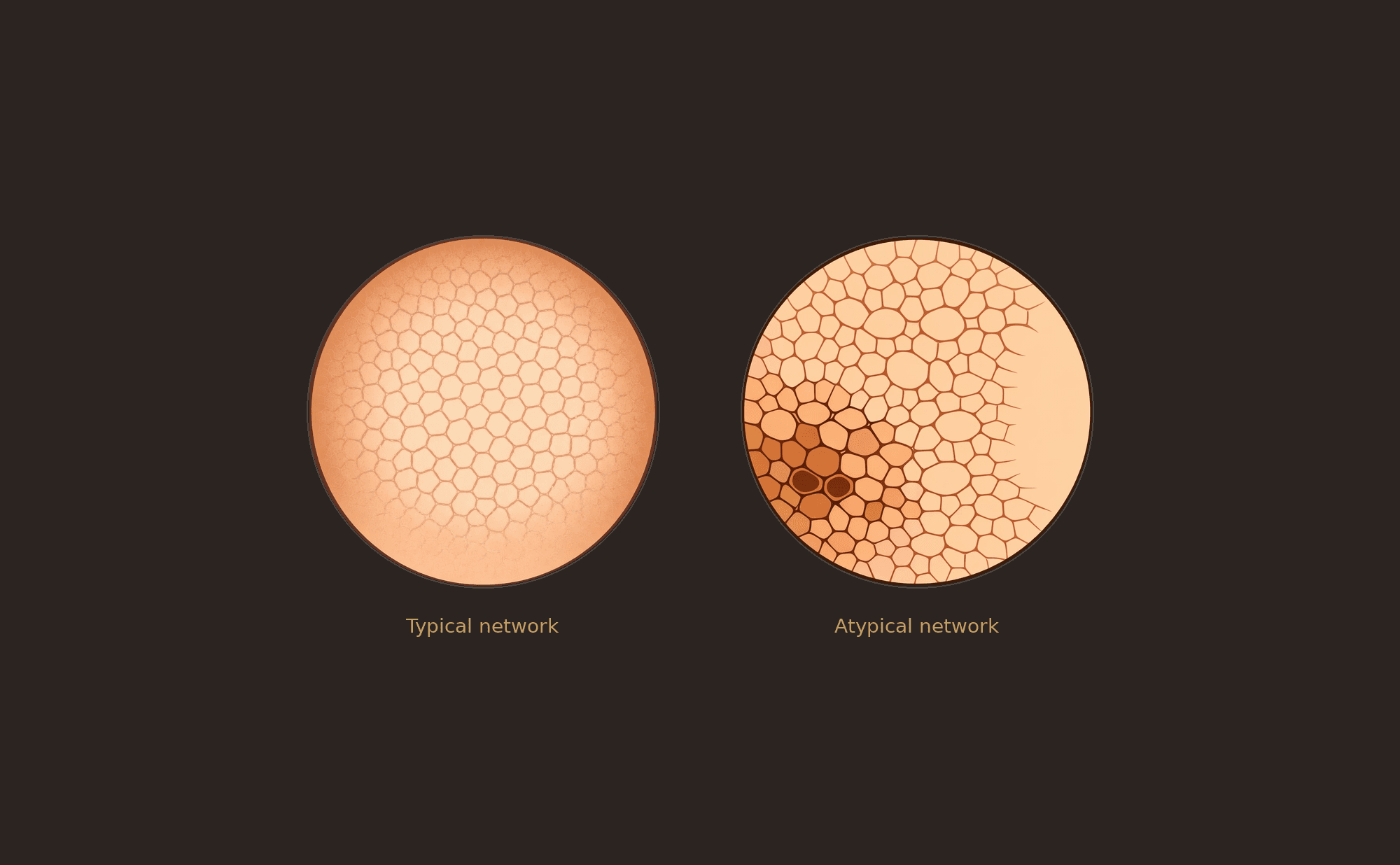
Left: typical network — even honeycomb mesh with uniform line thickness. Right: atypical network — irregular spacing, variable thickness, broken and branching lines
Associated with: Melanoma, atypical naevi. See Malignant Patterns.
B
Blue-grey globules
Plain language: Small round grey-blue dots — intermediate in size.
What you see: Well-defined round structures, grey-blue in colour. Intermediate in size between fine peppering granules and larger ovoid nests. May be scattered or clustered.
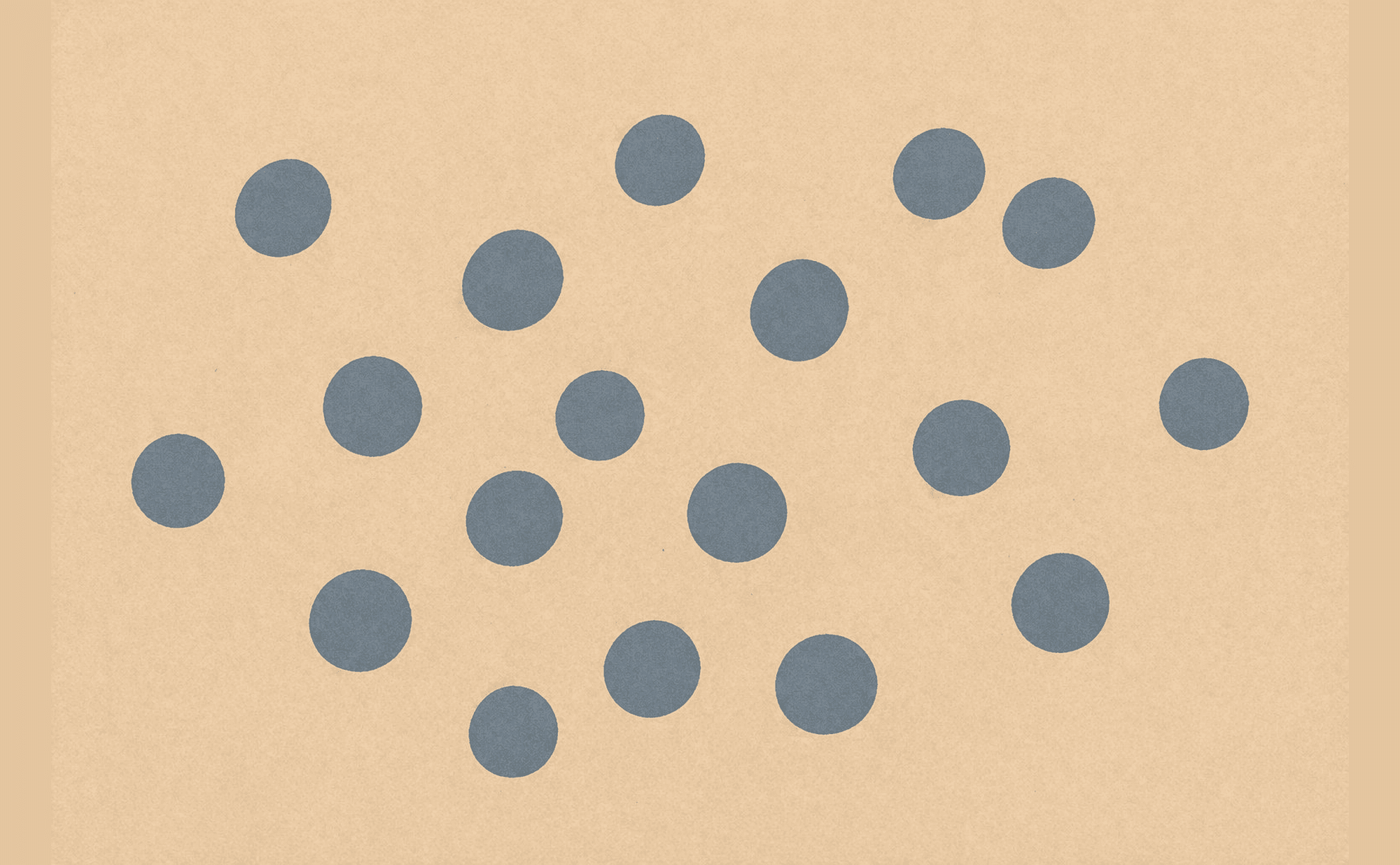
Blue-grey globules: well-defined round grey-blue structures — intermediate between fine peppering granules and larger ovoid nests
Associated with: Pigmented BCC. See Blue, Black & Grey and Non-Melanoma Skin Cancer.
Blue-grey ovoid nests
Plain language: Oval grey-blue structures with well-defined edges.
What you see: Discrete, egg-shaped grey-blue structures, clearly demarcated from surrounding tissue. Larger and more defined than globules. Individually countable.
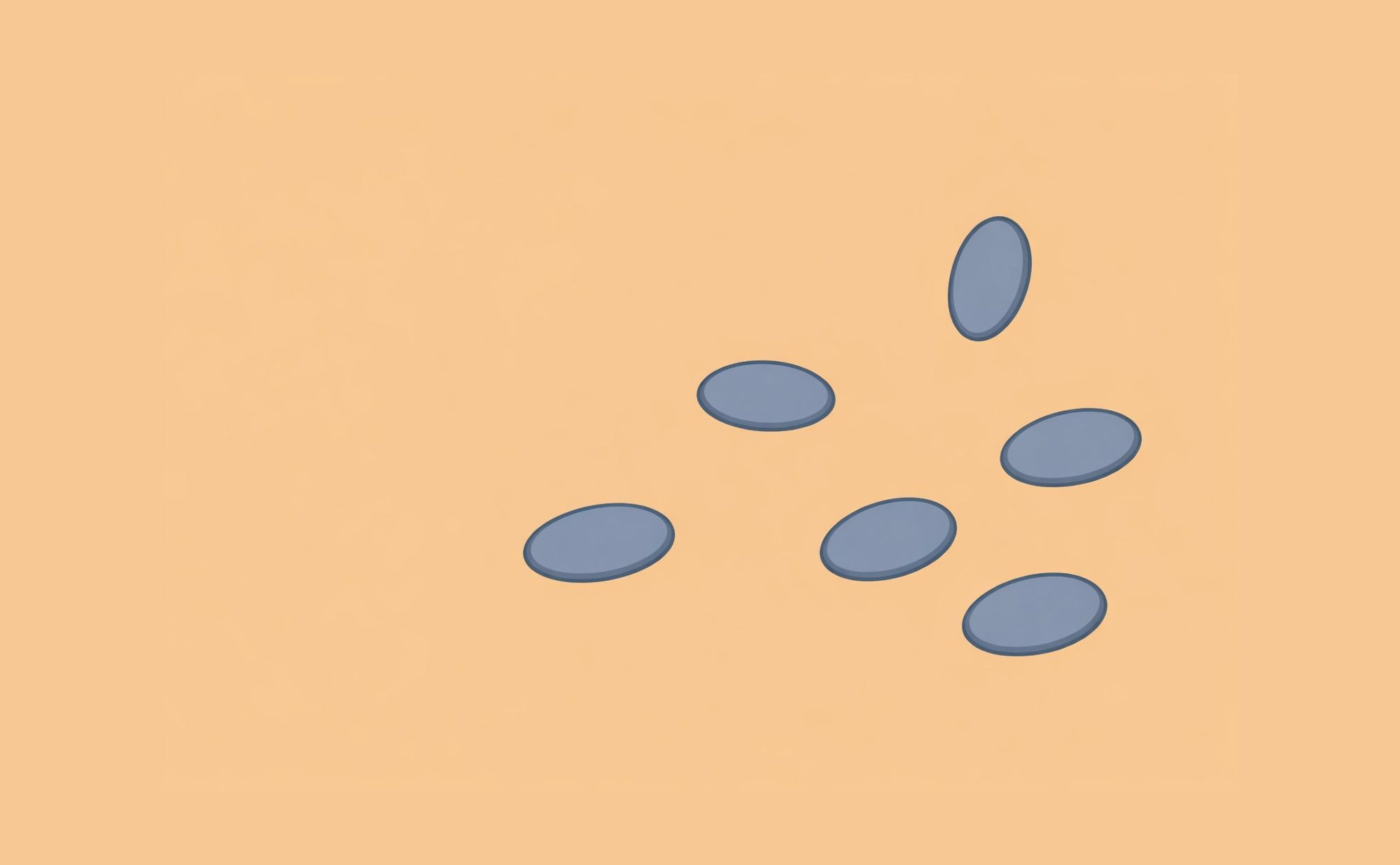
Blue-grey ovoid nests: discrete egg-shaped grey-blue structures — larger and more defined than globules, highly characteristic of pigmented BCC
Associated with: Pigmented BCC (highly characteristic). See Blue, Black & Grey and Non-Melanoma Skin Cancer.
Blue naevus
Plain language: A benign mole with deep blue colour.
What you see: Homogeneous steel-blue colour throughout — structureless and well-demarcated. The blue colour reflects melanin deep in the reticular dermis. Stable over time.

Blue naevus: homogeneous steel-blue colour, structureless and well-demarcated — melanin deep in the reticular dermis
Associated with: Benign melanocytic lesion. See Benign Patterns.
Blue-white veil
Plain language: Blue-grey colour with a whitish haze over it — like viewing blue through frosted glass.
What you see: Confluent blue-grey pigmentation covered by a ground-glass or frosted white overlay. Often over raised or palpable parts of a lesion. Both the blue and white components must be present.
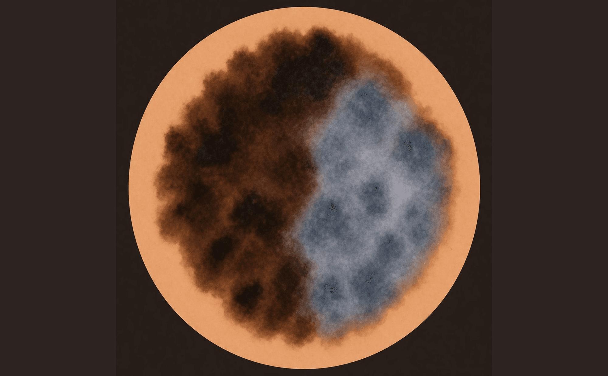
Blue-white veil: confluent blue-grey colour with frosted white overlay — both components must be present
Associated with: Melanoma (indicates dermal melanin with epidermal changes). See Blue, Black & Grey and Malignant Patterns.
C
Chrysalis structures
See Shiny white lines below. Chrysalis structures, crystalline structures, and shiny white lines are synonymous terms for the same feature.
Comedo-like openings
Plain language: Dark round or oval plugs resembling blackheads.
What you see: Round to oval, dark brown-black structures representing keratin plugs within follicular invaginations. A surface feature best seen with non-polarised dermoscopy.
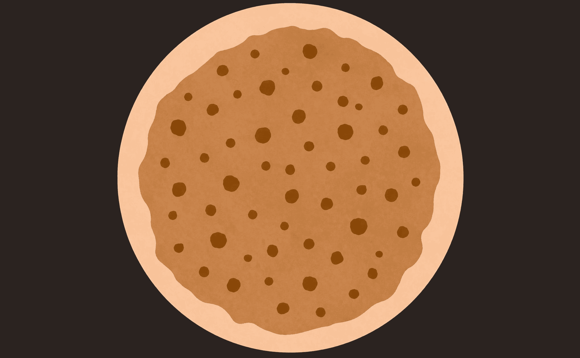
Comedo-like openings: dark round keratin plugs within the lesion surface — a hallmark of seborrhoeic keratosis
Associated with: Seborrhoeic keratosis (characteristic feature). See Benign Patterns.
Comma vessels
Plain language: Short, gently curved blood vessels shaped like commas.
What you see: Short curved vessels of uniform size, evenly distributed across the lesion. Slightly out of focus compared to surface vessels, indicating their depth in the dermis. Gently curved — not sharply angled or branching.
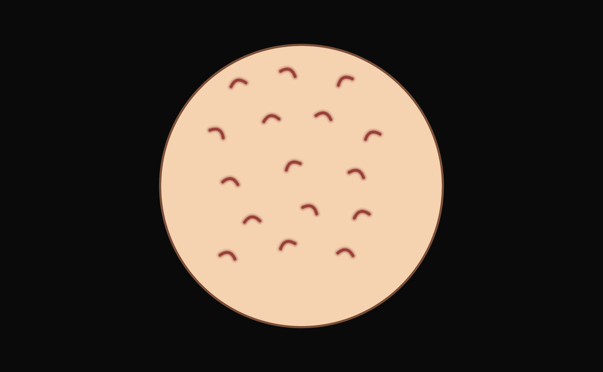
Comma vessels: short curved vessels of uniform size and even distribution — a reassuring feature of intradermal naevus
Associated with: Intradermal naevus (reassuring feature). See Vessels and Benign Patterns.
D
Dotted vessels
Plain language: Tiny red dots — blood vessels seen end-on.
What you see: Small red dots (like pinpricks) representing the tips of vertical capillary loops viewed from above. Significance depends entirely on context and distribution.
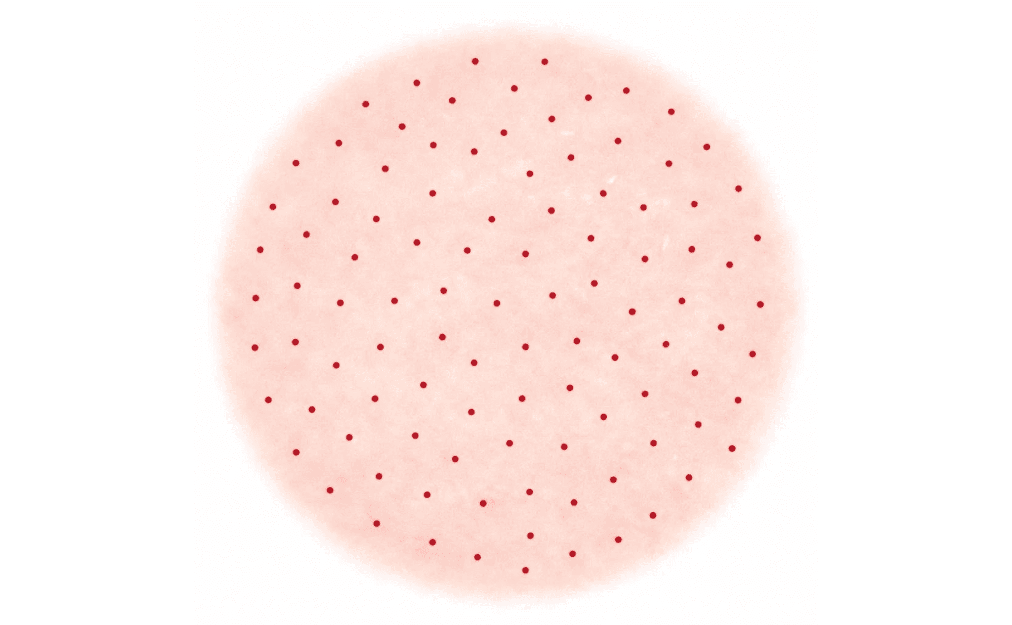
Dotted vessels: tiny red dots — capillary loops viewed end-on. Significance depends on context and distribution
Associated with: Various — Spitz naevus (regular pattern), melanoma (irregular pattern), Bowen's disease, inflammatory conditions. See Vessels.
F
Fissures and ridges
Plain language: Brain-like surface pattern with grooves and raised areas.
What you see: A cerebriform (brain-like) surface texture with linear depressions (fissures) between raised ridges. A surface feature reflecting the epidermal architecture of the lesion.
What you see
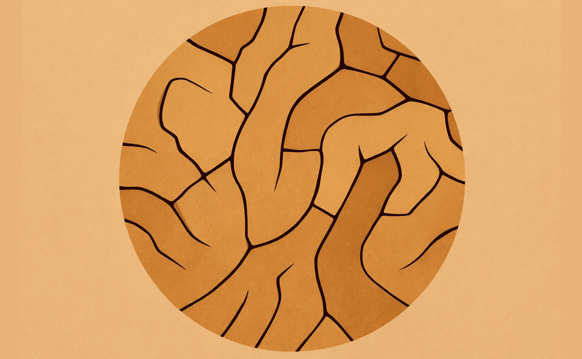
Fissures and ridges: cerebriform surface pattern — grooves and raised areas creating a brain-like texture characteristic of seborrhoeic keratosis
What it resembles
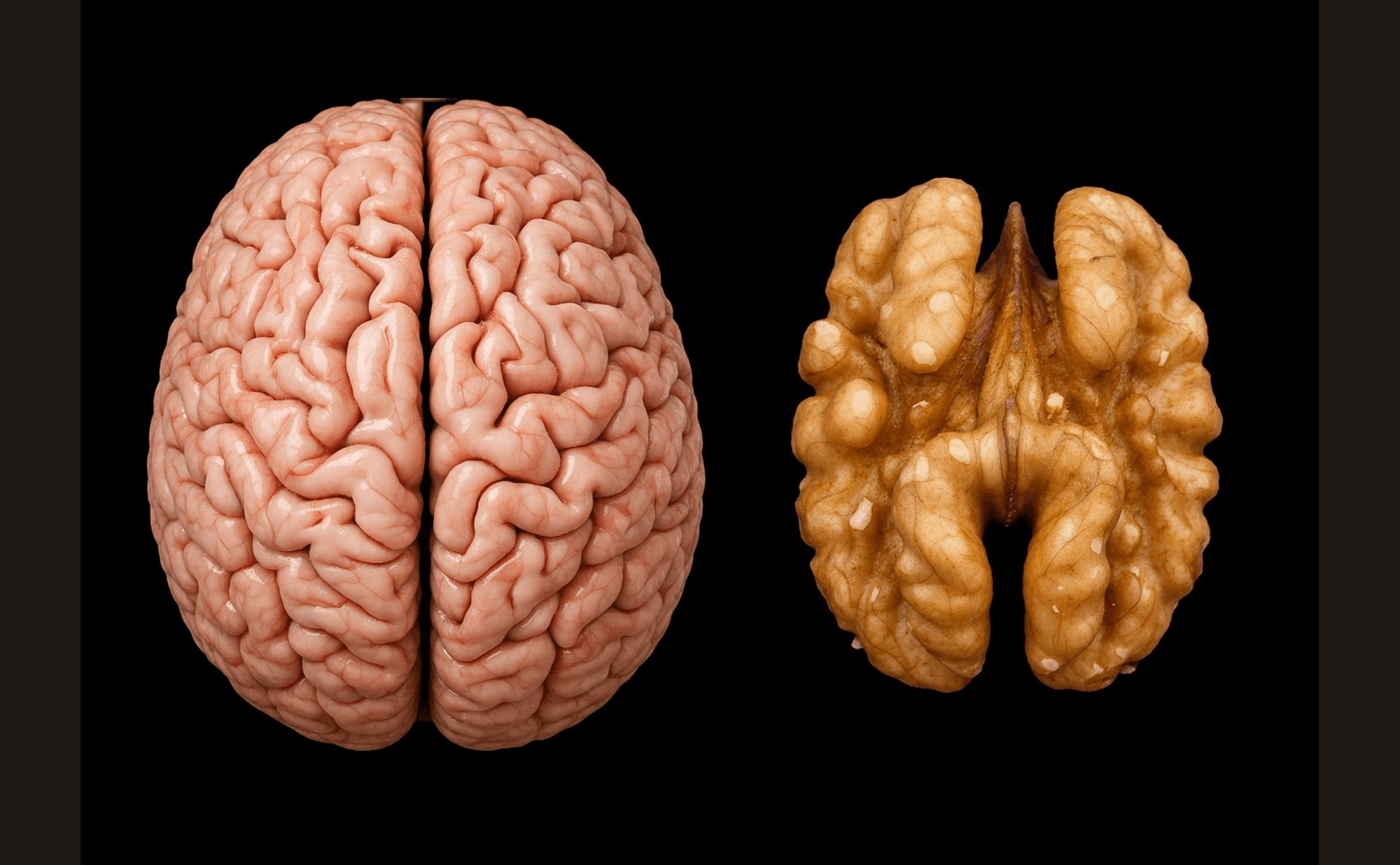
A walnut's wrinkled surface — the same cerebriform fold-and-groove pattern
Associated with: Seborrhoeic keratosis. See Benign Patterns.
G
Glomerular vessels
Plain language: Tightly coiled blood vessels resembling kidney glomeruli.
What you see: Small, tightly wound coiled vessels appearing as tangled red clusters. Often grouped together rather than evenly distributed.
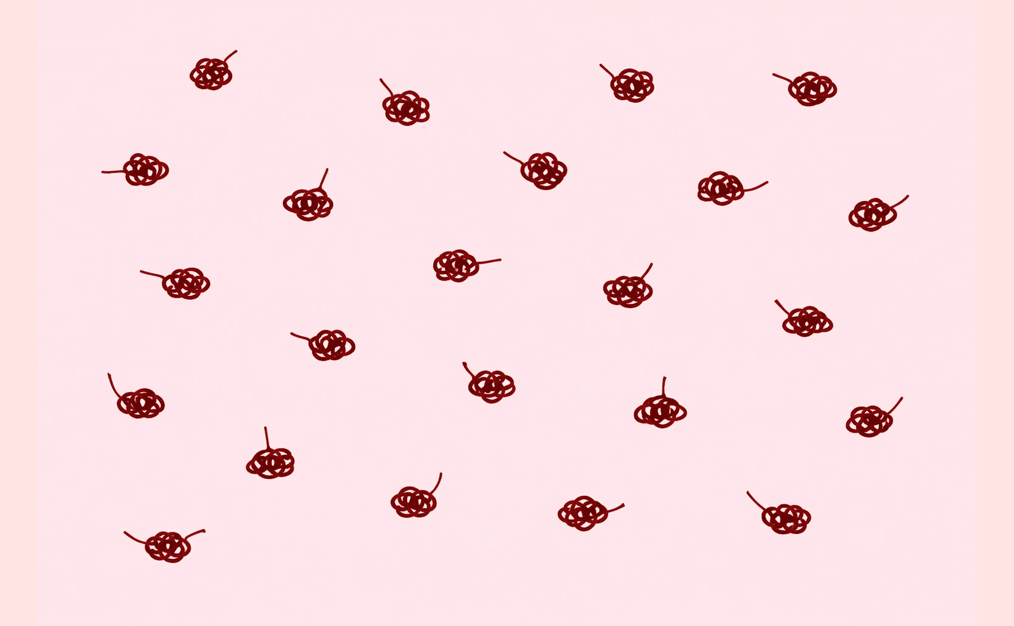
Glomerular vessels: tightly wound coiled vessels in tangled clusters — characteristic of Bowen's disease and SCC
Associated with: Bowen's disease (SCC in situ), invasive SCC. See Vessels and Non-Melanoma Skin Cancer.
Globules
Plain language: Round or oval brown structures — like small blobs of pigment.
What you see: Well-defined round to oval brown structures. When regular and uniformly distributed, they suggest a benign naevus (often a growing naevus in a young person). When irregular in size, shape, and distribution, they suggest melanoma.

Globules: round brown structures — regular and uniform in benign naevi, irregular in size and distribution when concerning
Associated with: Naevi (regular globules), melanoma (irregular globules). See Benign Patterns and Malignant Patterns.
H
Hairpin vessels
Plain language: U-shaped blood vessels — like hairpins or bobby pins.
What you see: Looped vessels forming U or hairpin shapes, often surrounded by a white keratinous halo. The halo indicates surface keratinisation and is characteristic of keratinising lesions.
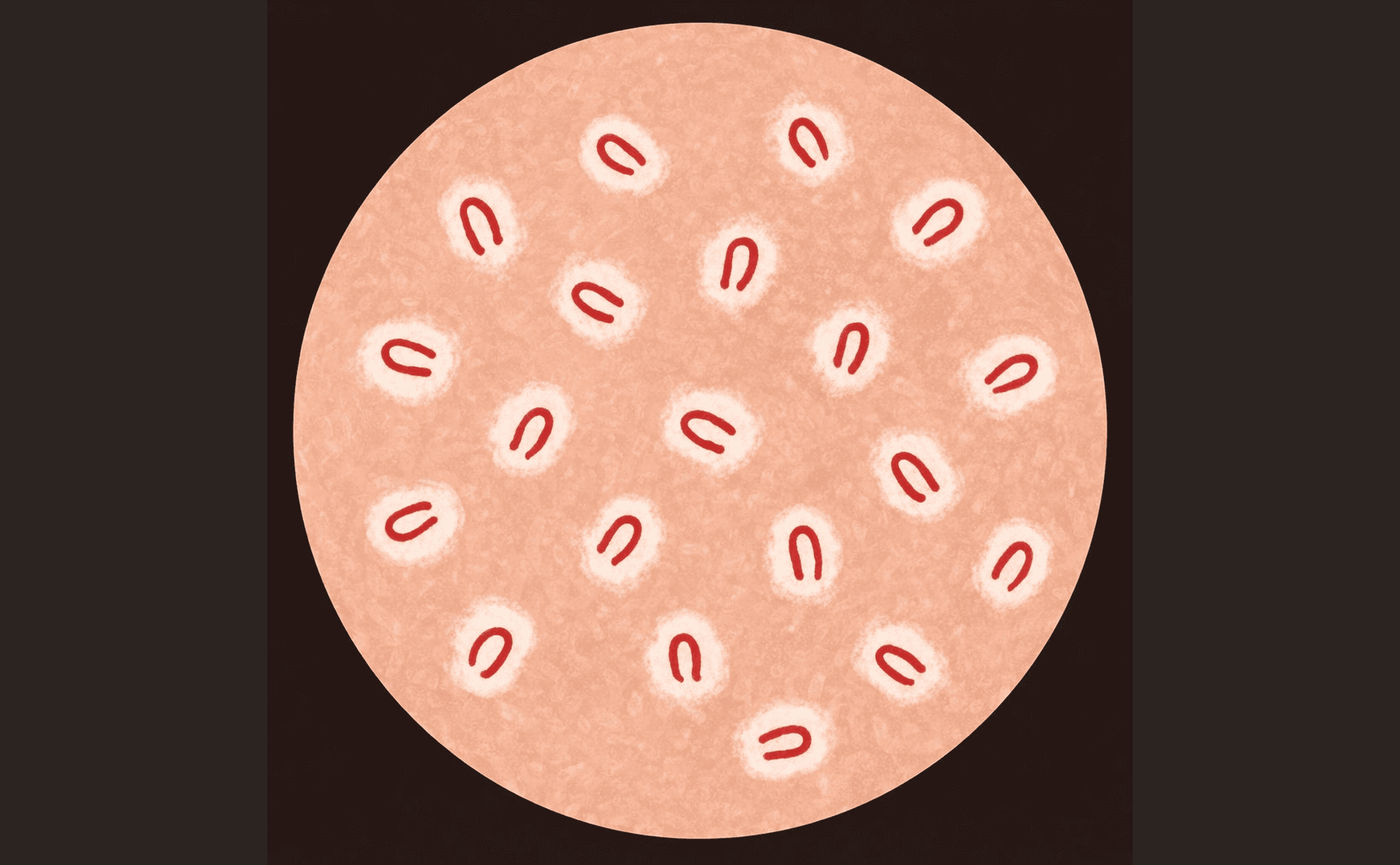
Hairpin vessels: U-shaped looped vessels with white keratinous halos — characteristic of keratinising lesions
Associated with: Seborrhoeic keratosis, keratoacanthoma, viral warts, SCC. See Vessels.
Homogeneous pattern
Plain language: Uniform colour throughout without distinct structures.
What you see: Even, structureless colour — brown, blue, tan, or pink — without network, globules, dots, or other discrete features. The absence of structure is itself a pattern.
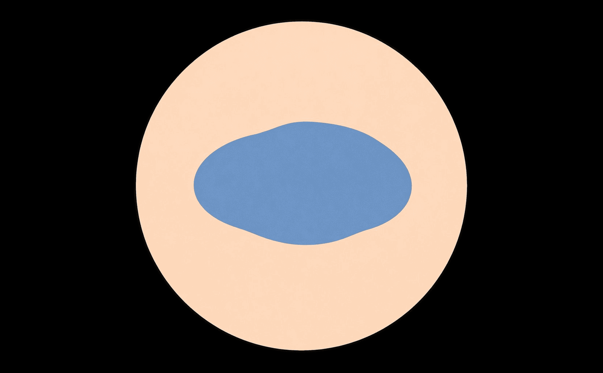
Homogeneous pattern: even, structureless colour — the absence of discrete features is itself a recognisable pattern
Associated with: Blue naevus (blue), intradermal naevus (tan or pink), some melanomas. See Benign Patterns.
I
Irregular dots and globules
Plain language: Dots and blobs of varying sizes scattered unevenly.
What you see: Round structures that vary in size, shape, and distribution — lacking the uniformity and symmetry of benign globules. Scattered asymmetrically across the lesion.
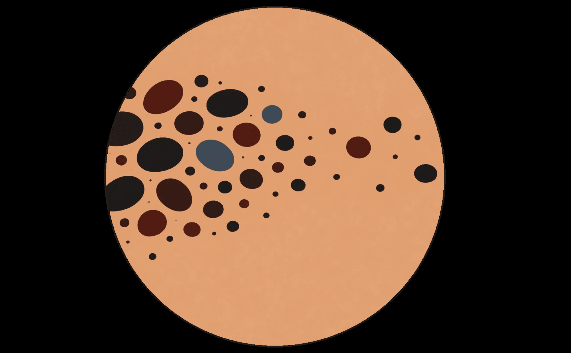
Irregular dots and globules: varying sizes scattered asymmetrically — lacking the uniformity of benign globules
Associated with: Melanoma. See Malignant Patterns.
Irregular streaks
Plain language: Finger-like projections at the edges that are not symmetrically distributed.
What you see: Radial projections at the lesion periphery that vary in length, thickness, and distribution. Asymmetric — present in some areas and absent in others. Represent confluent junctional nests of melanocytes extending outward.
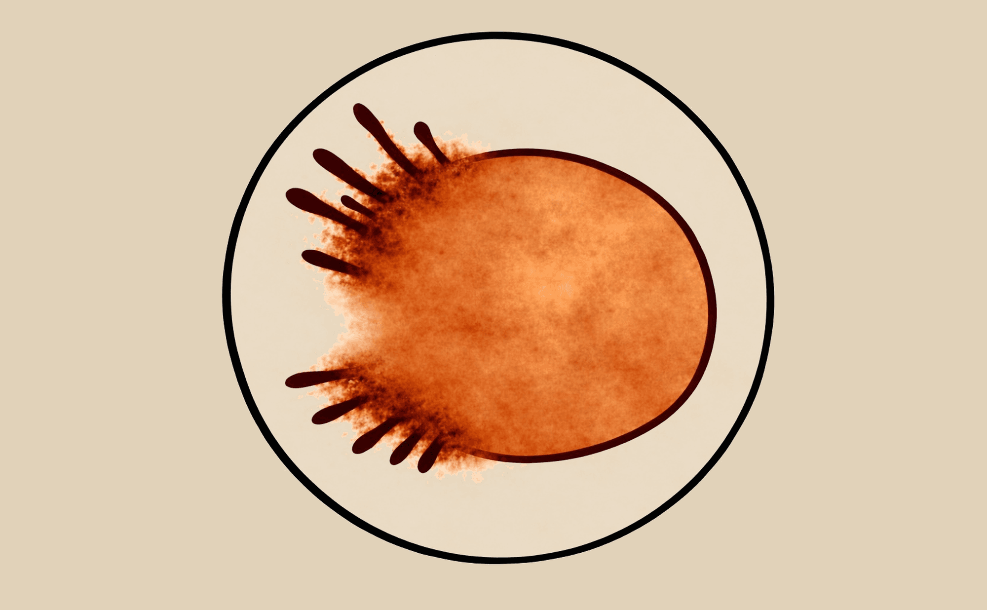
Irregular streaks: asymmetric radial projections at the periphery — variable length, thickness, and distribution
Associated with: Melanoma. Contrast with the symmetric starburst pattern of Spitz naevus. See Malignant Patterns.
L
Lacunae
Plain language: Round, blood-filled spaces separated by pale walls — like a stained glass window.
What you see: Well-defined round or oval red-purple areas representing dilated vascular spaces, separated by pale septa (walls). The compartmentalised architecture is the defining feature.
What you see
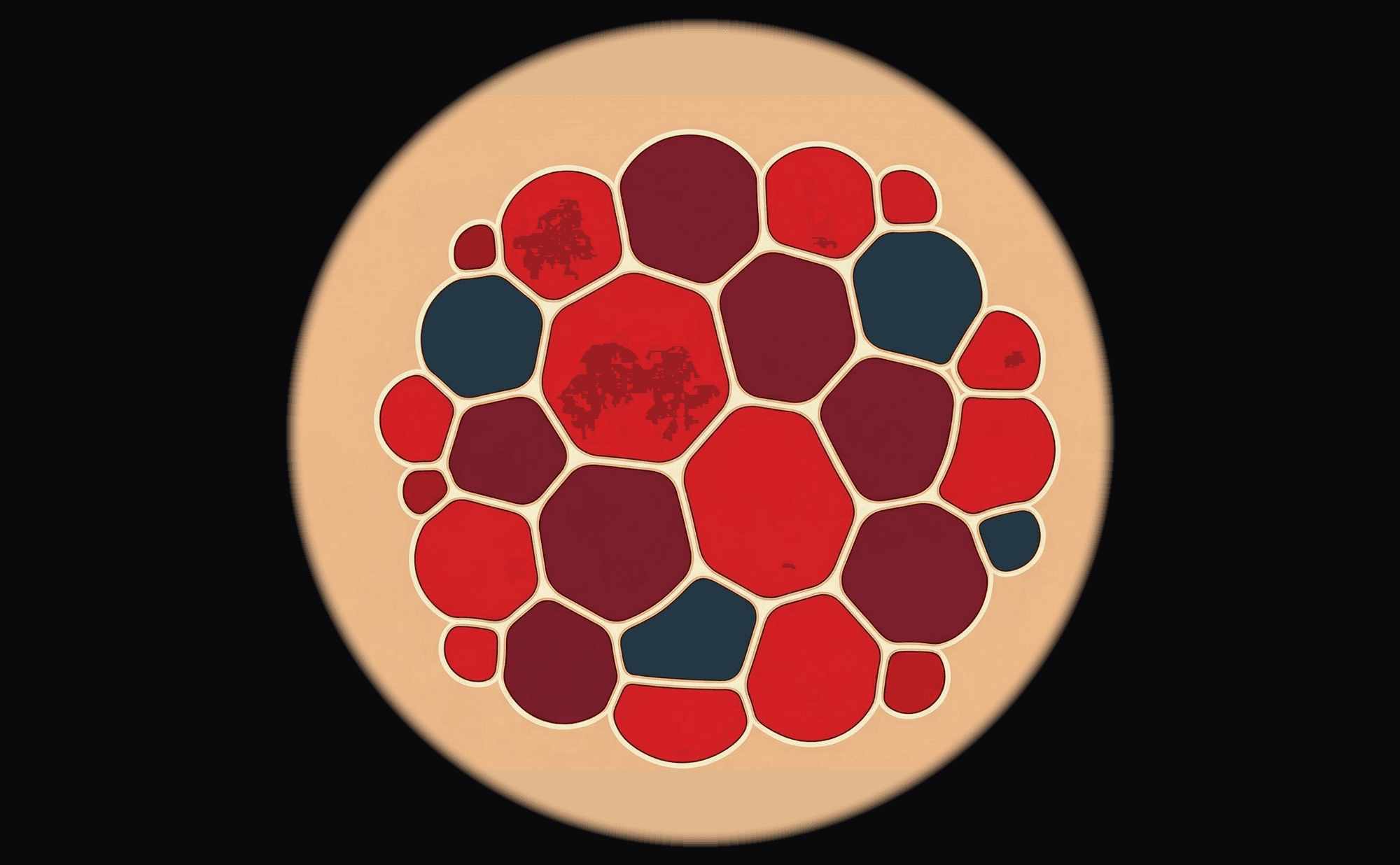
Lacunae: round red-purple vascular compartments separated by pale septa — the hallmark stained-glass-window pattern
What it resembles
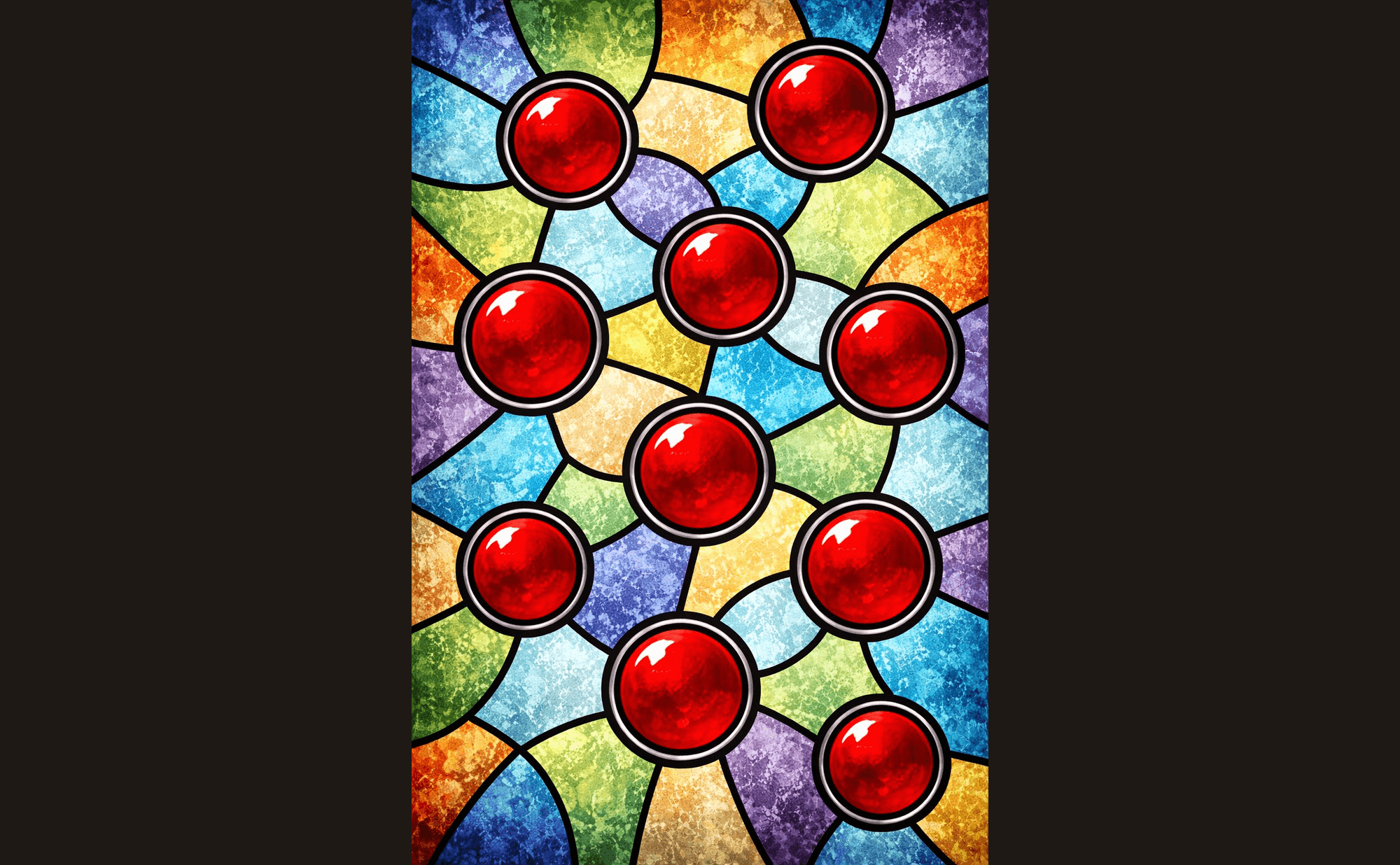
Stained glass — round coloured compartments with dividing walls, exactly like the lacunar pattern
Associated with: Cherry angioma (characteristic), angiokeratoma. See Benign Patterns and Vessels.
Leaf-like areas
Plain language: Brown-grey structures with finger-like projections resembling maple leaves.
What you see: Pigmented structures at the lesion periphery with bulbous, finger-like projections. Connected to the lesion body, not free-floating.
What you see
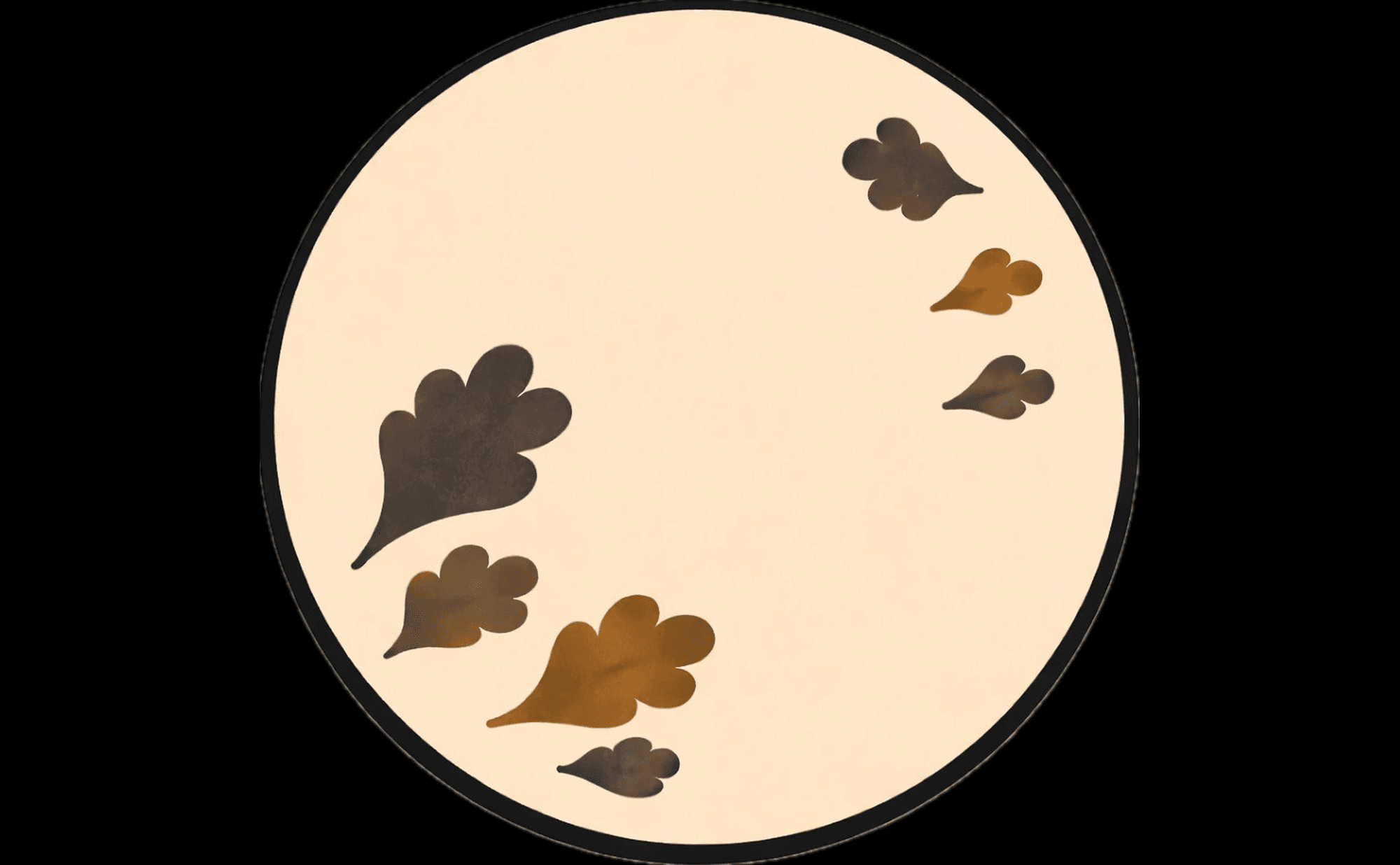
Leaf-like areas: brown-grey structures with bulbous finger-like projections at the periphery — highly specific for pigmented BCC
What it resembles

A leaf's finger-like projections — the visual origin of the term 'leaf-like areas'
Associated with: Pigmented BCC (highly specific). See Non-Melanoma Skin Cancer and Blue, Black & Grey.
M
Milia-like cysts
Plain language: Small white or yellowish round structures — like tiny pearls embedded in the surface.
What you see: Discrete, round, bright white-to-yellowish structures representing intraepidermal horn cysts. Best seen with non-polarised dermoscopy.
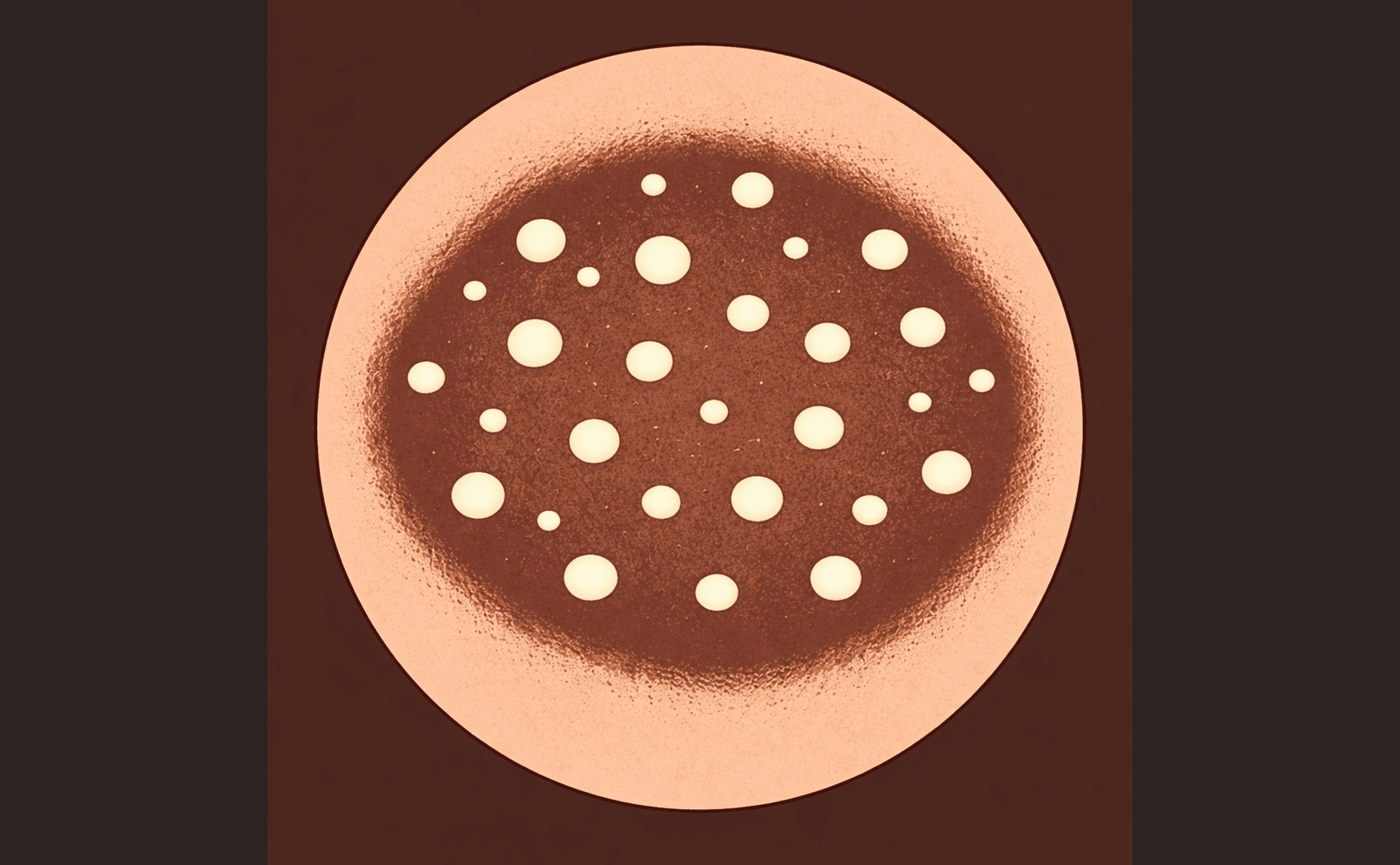
Milia-like cysts: bright white-yellow round structures like tiny pearls — a hallmark of seborrhoeic keratosis
Associated with: Seborrhoeic keratosis (characteristic feature). See Benign Patterns.
Milky-red areas
Plain language: Hazy, diffuse pink-red colour — not individual vessels but a blush.
What you see: Structureless pink-red zones with a hazy, milky quality. Individual vessels cannot be resolved within these areas — the redness is diffuse, like a vascular blush seen through frosted glass.
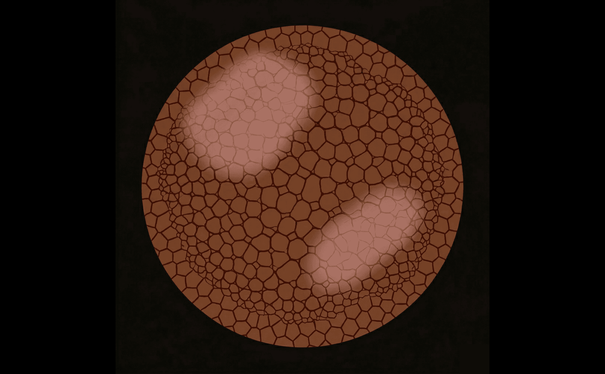
Milky-red areas: diffuse pink-red zones with a hazy quality — individual vessels cannot be resolved within the blush
Associated with: Melanoma (especially invasive or amelanotic), Spitz naevi. See Vessels and Malignant Patterns.
N
Negative network
Plain language: White or pale lines forming a network pattern — the inverse of normal pigment network.
What you see: A grid of pale or white lines surrounding darker pigmented areas. The structural inverse of typical pigment network — light lines, dark spaces (rather than the usual dark lines, light spaces).
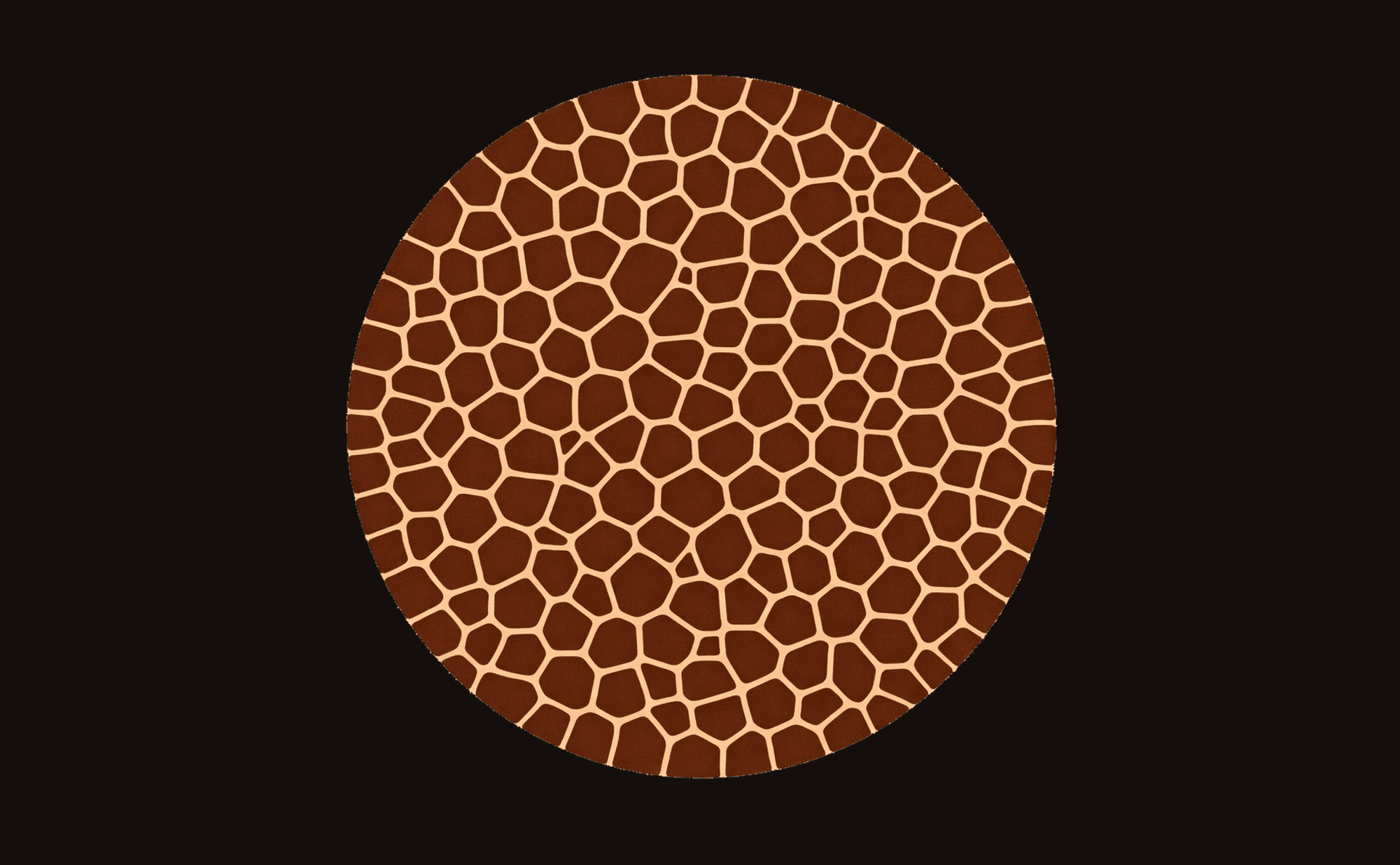
Negative network: pale lines surrounding darker spaces — the structural inverse of typical pigment network
Associated with: Melanoma, Spitz naevus. Concerning in adults. See Shiny White Structures.
P
Peppering
Plain language: Fine grey-blue dots scattered like ground pepper — a sign of immune attack on the lesion.
What you see: Fine grey-blue granules scattered across part of a lesion, representing melanin within melanophages (pigment-containing immune cells). Often accompanied by white scar-like areas. One of the seven TADA high-risk features.
What you see
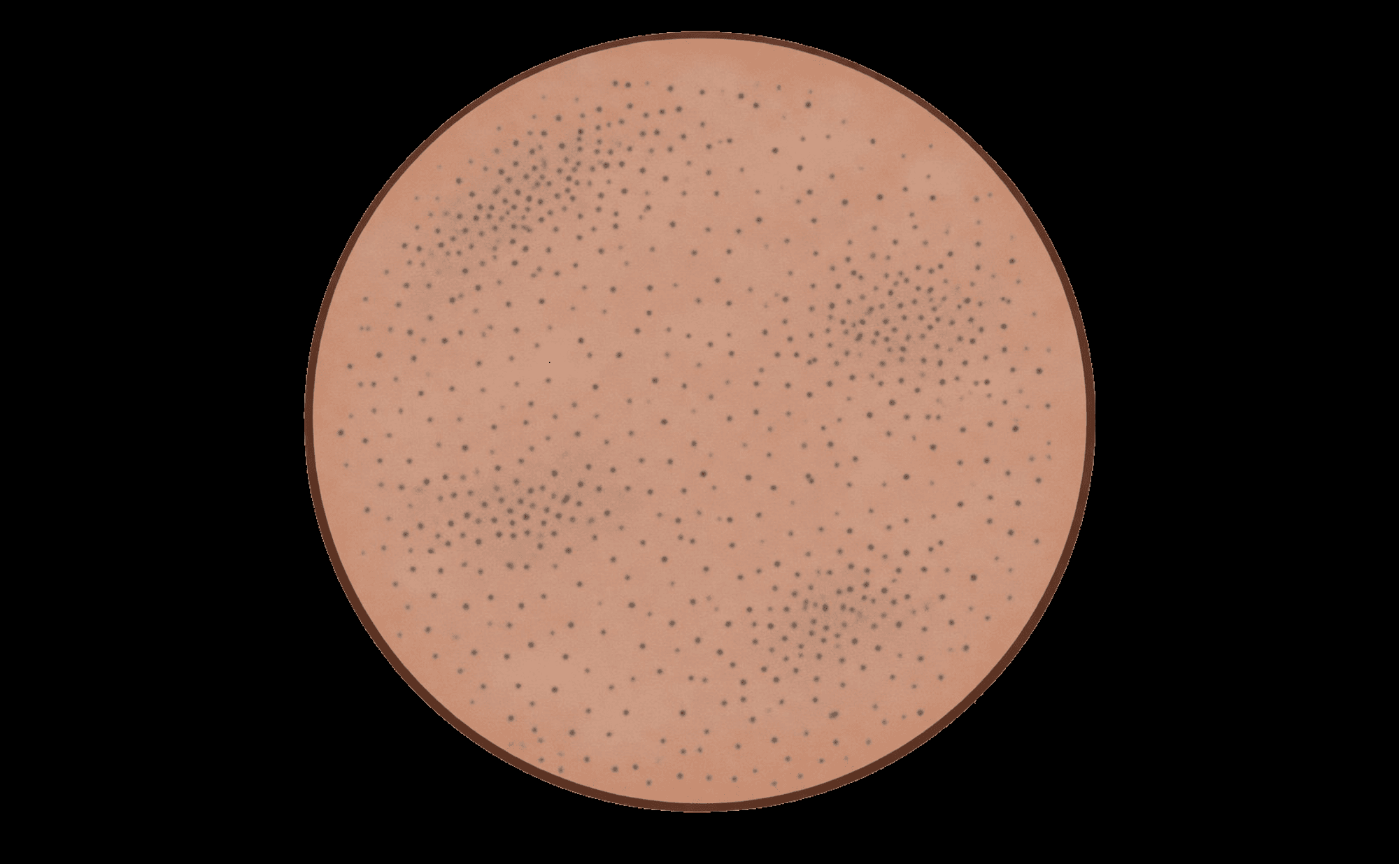
Peppering: fine grey-blue granules representing melanophages — one of the seven TADA high-risk features
What it resembles

Ground pepper scattered across a surface — the visual origin of the term
Associated with: Melanoma with regression, lichen planus-like keratosis. See Blue, Black & Grey.
Pigment network
Plain language: Brown honeycomb or mesh pattern — the most fundamental melanocytic structure.
What you see: A grid-like pattern of brown lines surrounding lighter spaces, representing melanin at the dermo-epidermal junction concentrated along the rete ridges. When regular (uniform mesh, even line thickness), it suggests a benign naevus. When atypical (irregular mesh, variable thickness), it raises concern.
What you see
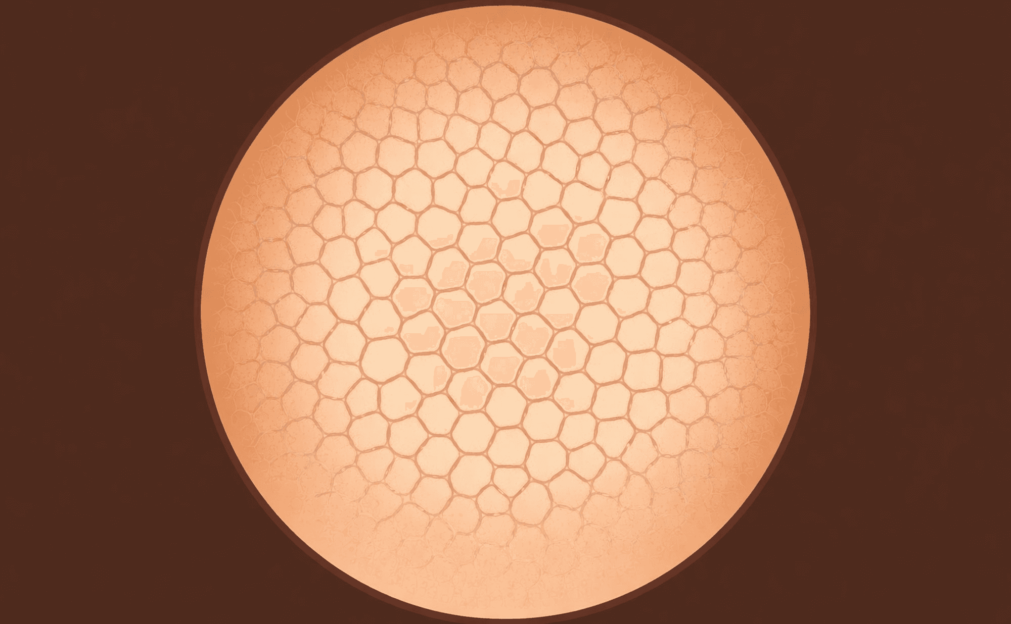
Typical pigment network: regular honeycomb mesh with uniform line thickness and gradual peripheral fading — the gold standard of normal melanocytic pattern
What it resembles
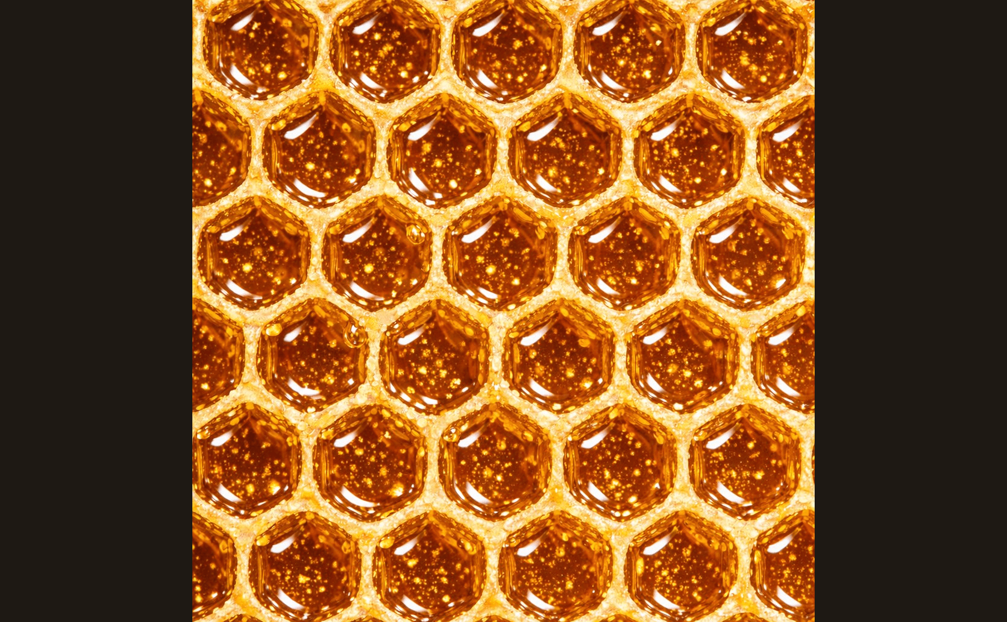
A honeycomb's regular hexagonal mesh — the classic visual parallel for a typical pigment network
Associated with: Melanocytic lesions — naevi and melanoma. Pattern regularity determines significance. See Benign Patterns and Malignant Patterns.
Polymorphous vessels
Plain language: Multiple different types of blood vessels coexisting within one lesion.
What you see: Two or more distinct vessel morphologies (e.g., dotted mixed with linear-irregular mixed with serpentine) in a single lesion, with chaotic distribution. The variety and disorder are the defining features. One of the seven TADA high-risk features.
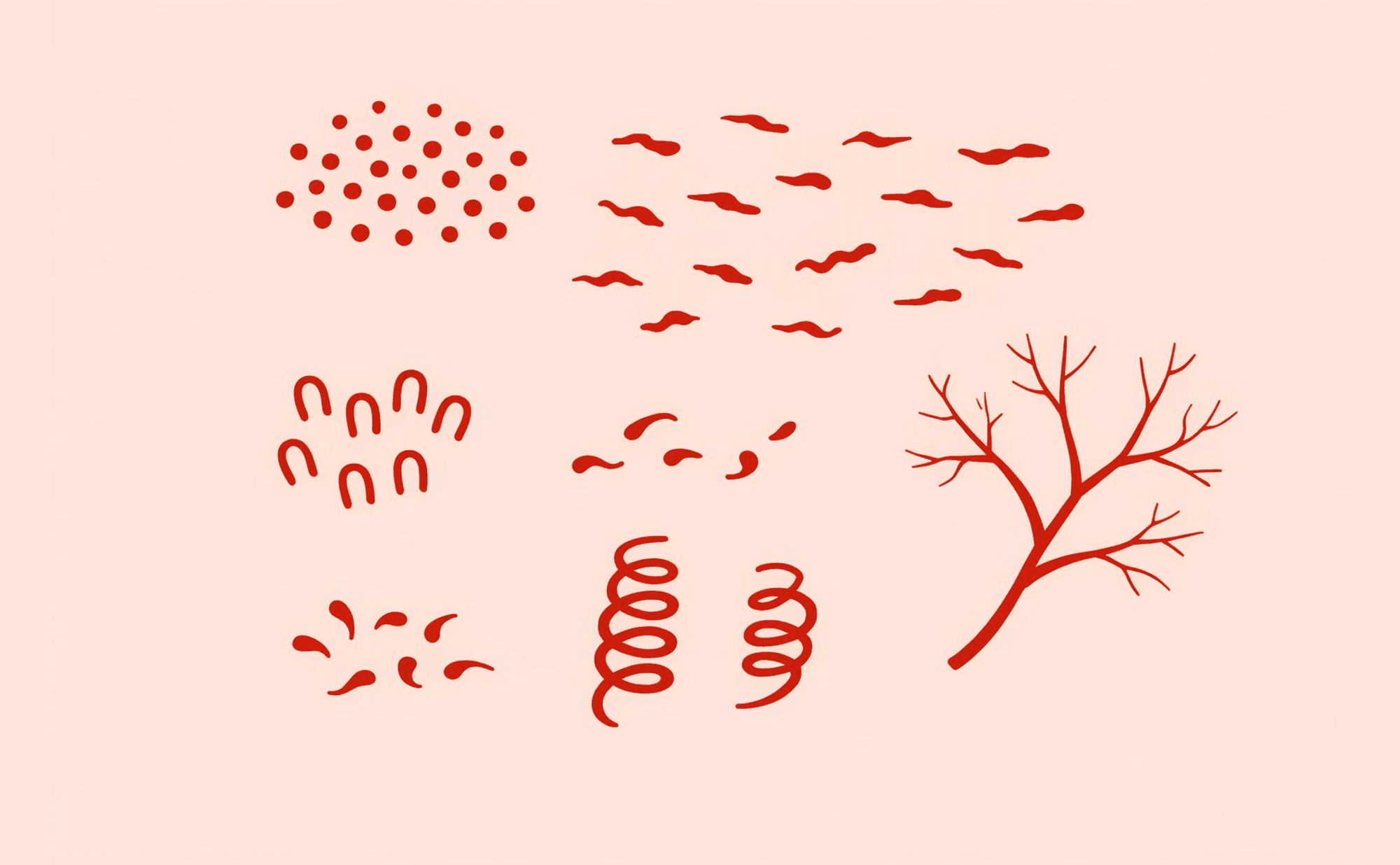
Polymorphous vessels: multiple vessel morphologies in chaotic distribution — the variety and disorder are highly concerning
Associated with: Melanoma (highly concerning). See Vessels and Malignant Patterns.
R
Regression structures
Plain language: White scar-like areas with grey dots — indicating the body has attacked the lesion.
What you see: A combination of white structureless depigmentation (fibrosis) and grey peppering (melanophages) where the immune system has destroyed pigmented cells. Partial regression does not mean the threat is gone.
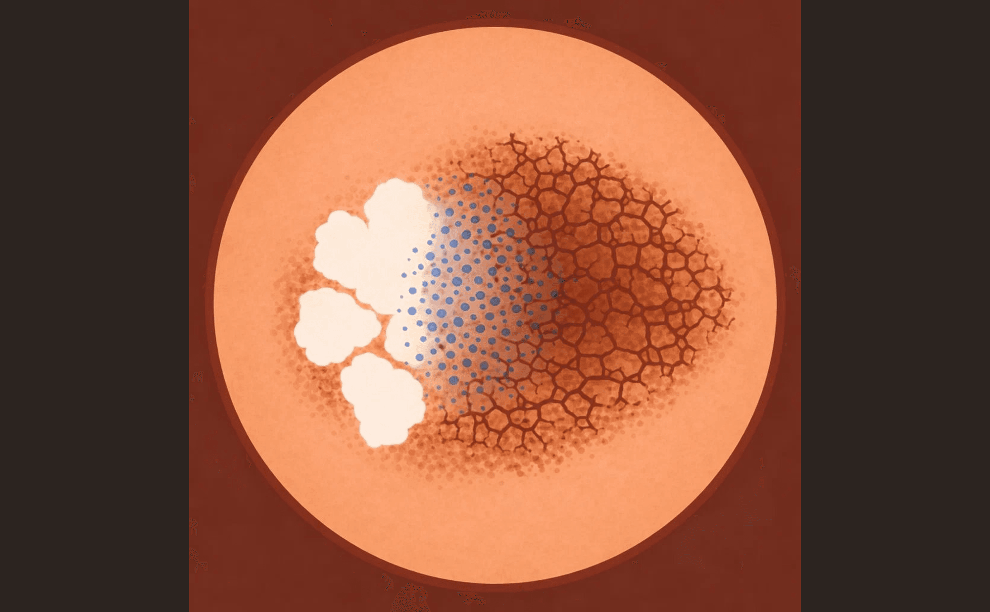
Regression structures: white scar-like fibrosis combined with grey-blue peppering — a three-zone gradient from white patches through peppering to disrupted network
Associated with: Melanoma with regression. See Blue, Black & Grey and Malignant Patterns.
Rhomboidal structures
Plain language: Grey-brown lines forming angular, diamond-like shapes around follicles.
What you see: Pigmented lines forming rhomboid (diamond or parallelogram) shapes, typically between follicular openings on sun-damaged facial skin. The angular geometry is distinctive.
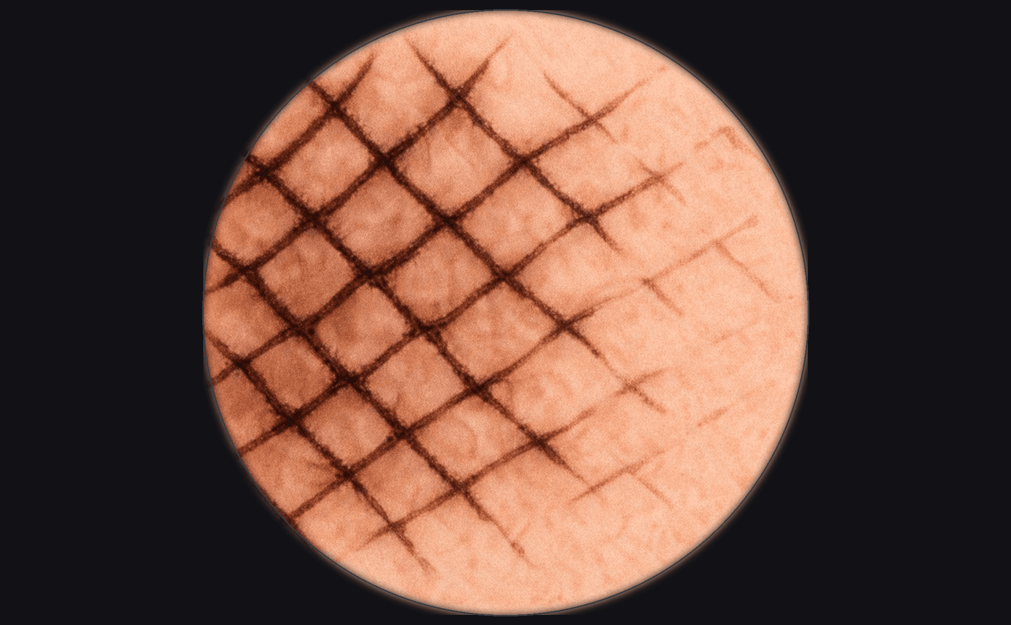
Rhomboidal structures: grey-brown lines forming diamond shapes between follicular openings — characteristic of lentigo maligna
Associated with: Lentigo maligna. See Blue, Black & Grey and Malignant Patterns.
Rosettes
Plain language: Four white dots arranged in a clover-leaf pattern (polarised light only).
What you see: Small four-dot structures resembling a four-leaf clover, visible only under polarised dermoscopy. Represent light interaction with concentric keratinisation around follicular openings.
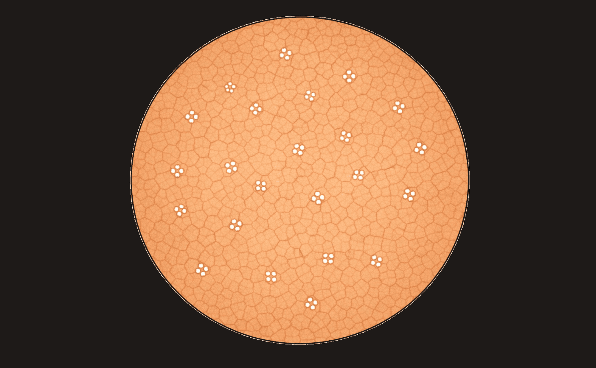
Rosettes: four white dots in clover-leaf arrangement — a polarised light feature associated with actinic keratosis and sun-damaged skin
Associated with: Actinic keratosis, sun-damaged skin. See Shiny White Structures and Non-Melanoma Skin Cancer.
S
Shiny white lines
Plain language: Bright white lines visible only under polarised light — also called chrysalis or crystalline structures.
What you see: Linear white structures, often crossing at angles, with a shiny or glistening quality. Only visible with polarised dermoscopy. Represent dermal collagen (fibrosis). One of the seven TADA high-risk features in melanocytic lesions.
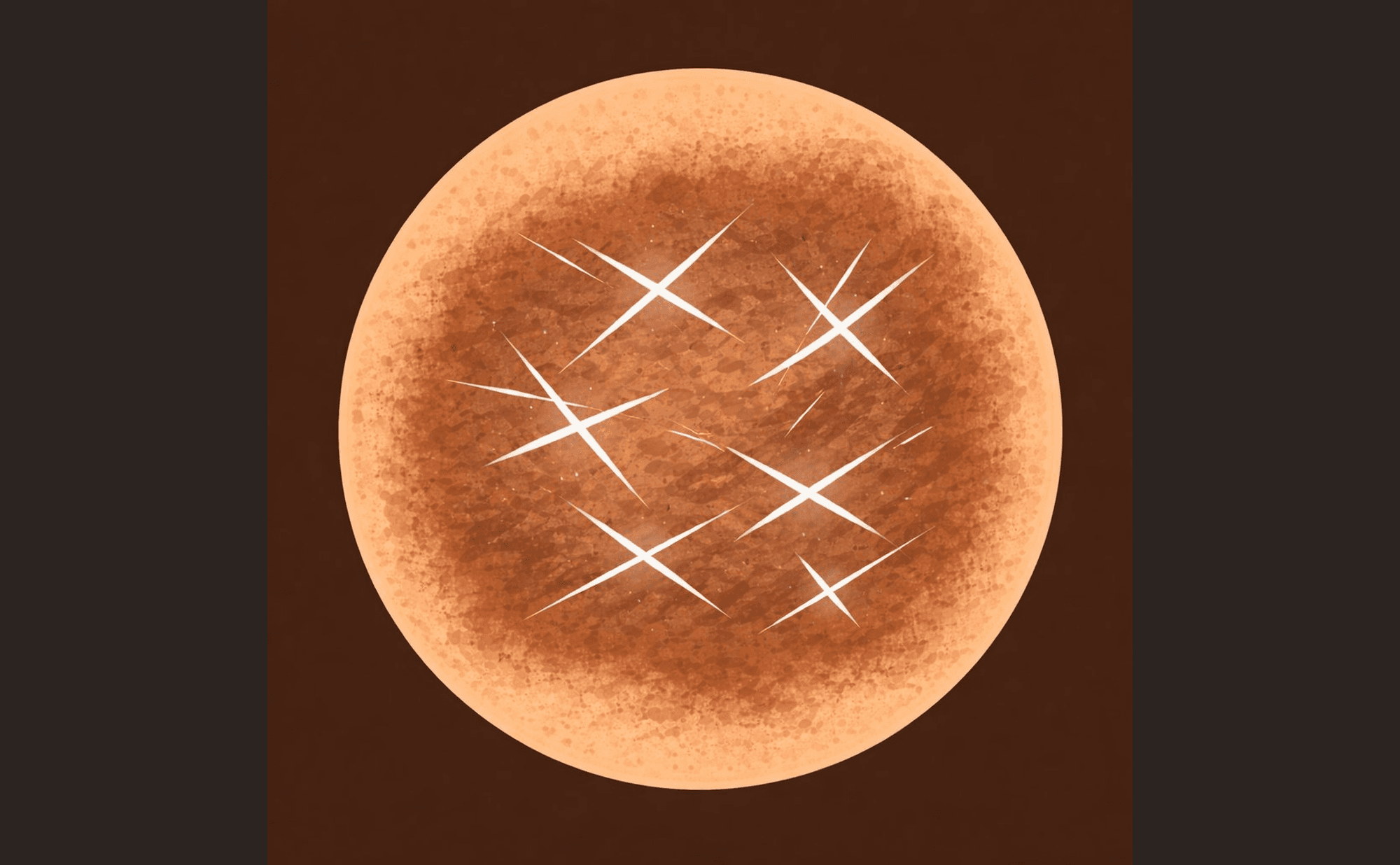
Shiny white lines: bright linear structures crossing at angles — visible only with polarised dermoscopy, representing dermal collagen
Associated with: Melanoma (indicates dermal involvement), BCC, dermatofibroma. See Shiny White Structures.
Shiny white blotches
Plain language: Bright white patches visible only under polarised light — larger than individual lines.
What you see: Broader areas of bright white colour under polarised light, representing the stromal collagen response. Larger and more confluent than shiny white lines.
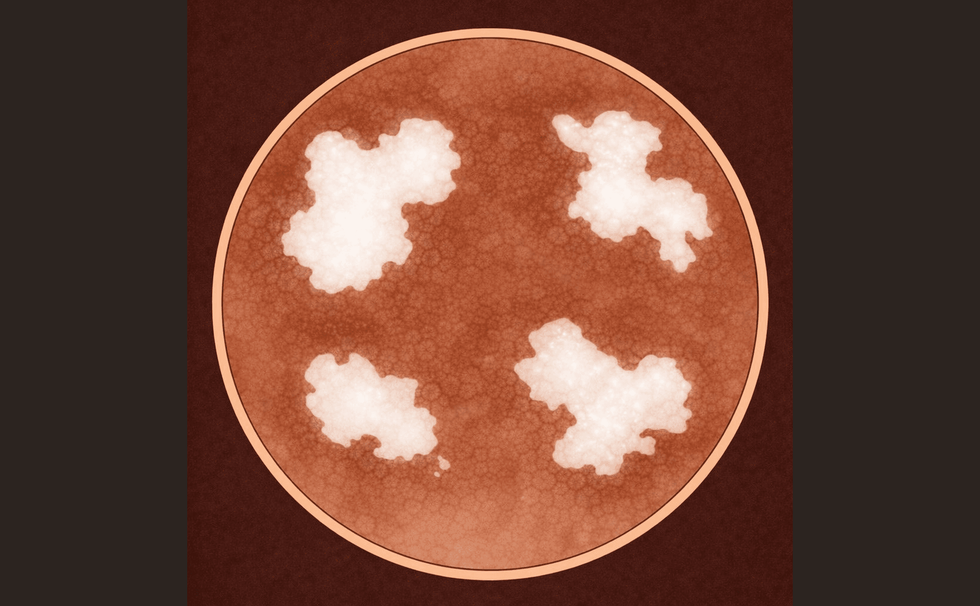
Shiny white blotches: broader bright white patches — representing stromal collagen response, characteristic of BCC
Associated with: BCC (characteristic feature). See Shiny White Structures and Non-Melanoma Skin Cancer.
Spoke-wheel structures
Plain language: Lines radiating toward a central darker point — like spokes of a bicycle wheel.
What you see: Radial brown-grey projections converging on a central hub, creating a wheel-spoke configuration.
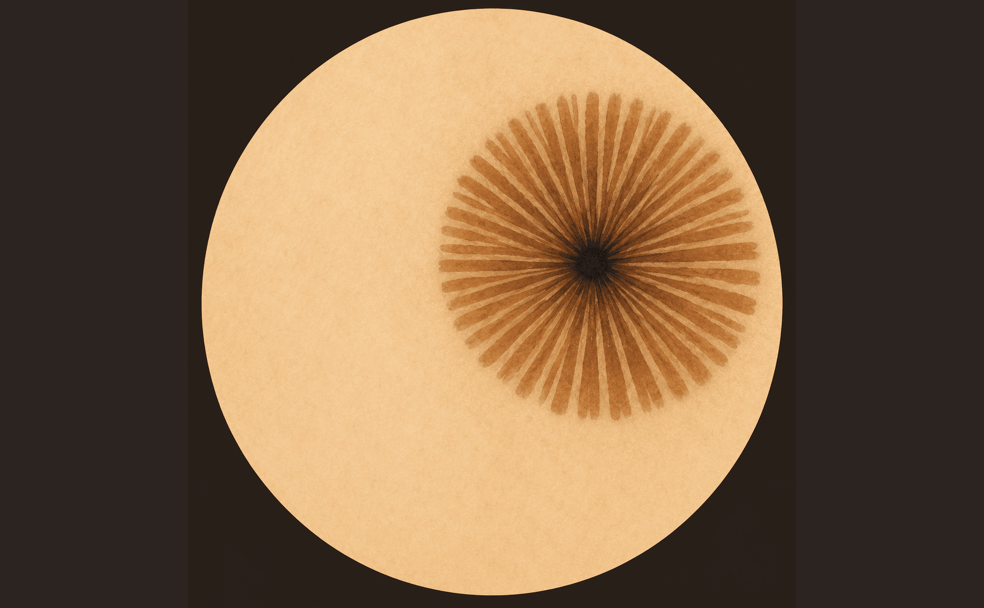
Spoke-wheel structures: radial projections converging on a central hub — a wheel-spoke configuration characteristic of pigmented BCC
Associated with: Pigmented BCC. See Blue, Black & Grey and Non-Melanoma Skin Cancer.
Starburst pattern
Plain language: Streaks or projections radiating symmetrically around the entire edge of a lesion.
What you see: Regular radial projections distributed symmetrically around the entire lesion periphery, creating a star-like or firework pattern. The symmetry is the key feature — asymmetric streaks are a different finding.
What you see
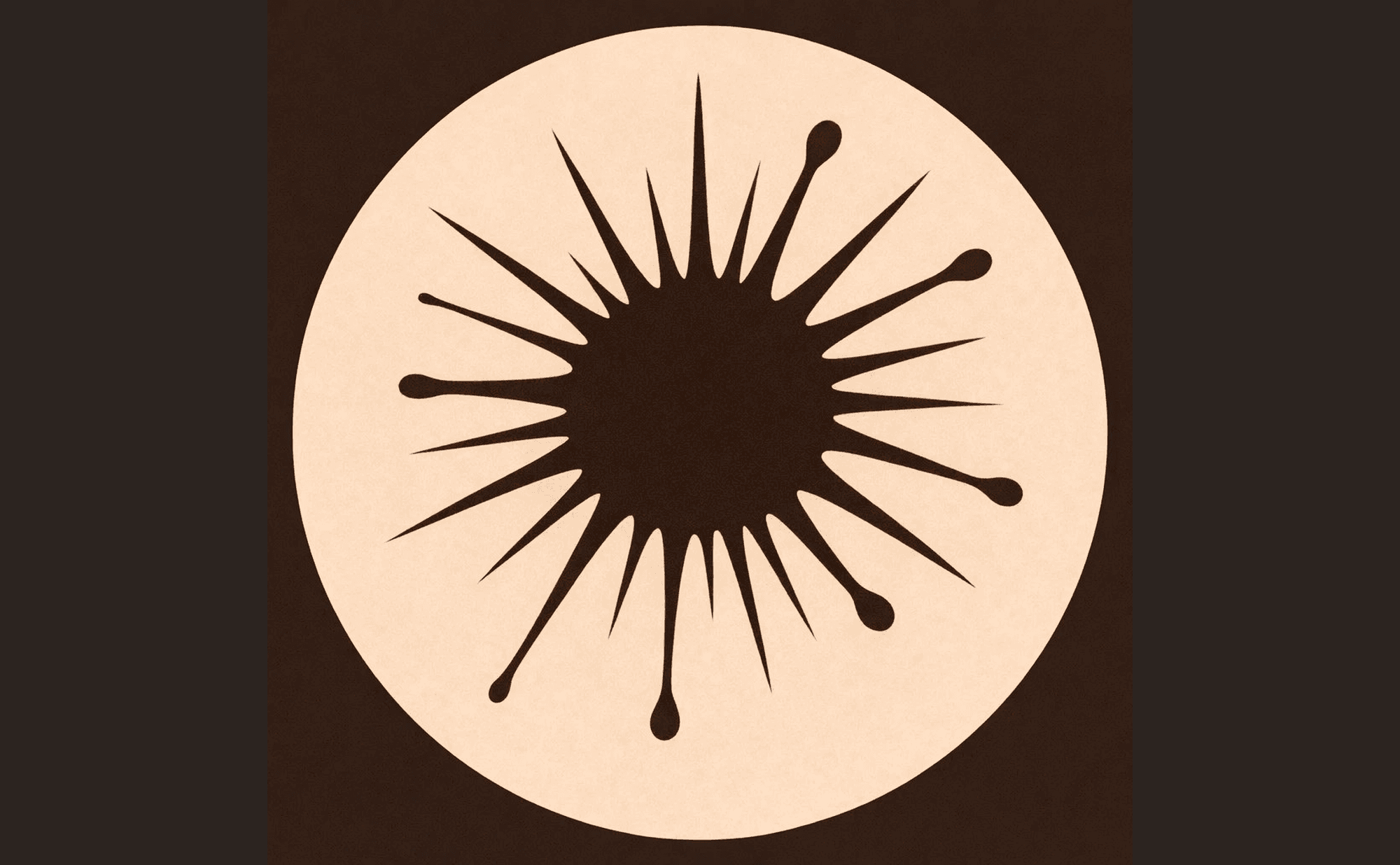
Starburst pattern: symmetric radial projections around the entire periphery — the symmetry distinguishes this from concerning irregular streaks
What it resembles
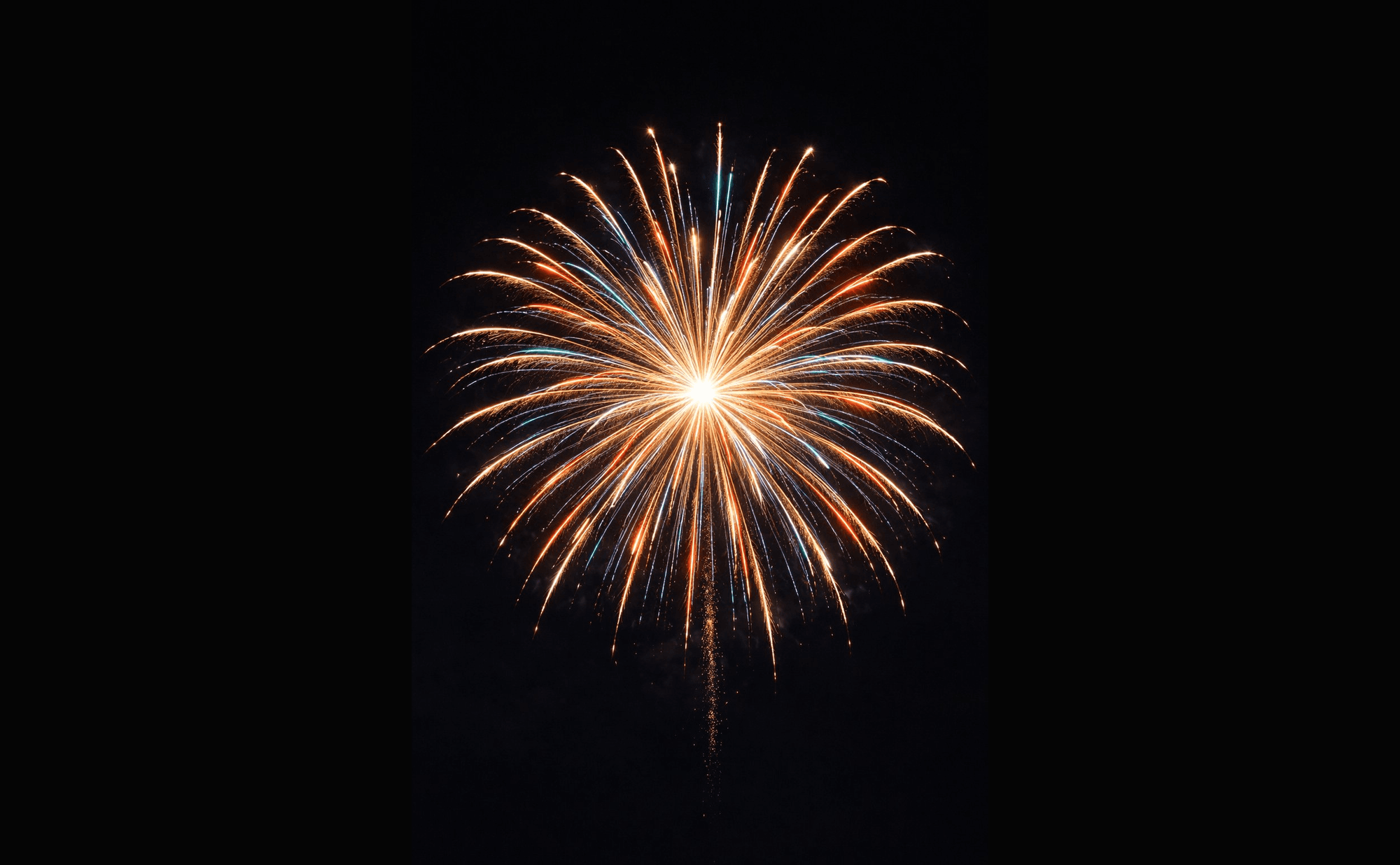
A firework's radial burst — symmetric streaks projecting outward from a central point
Associated with: Spitz or Reed naevus (when symmetric), melanoma (when asymmetric). See Benign Patterns.
Strawberry pattern
Plain language: Red background with white or yellow dots filling the pores.
What you see: An erythematous (pink-red) background with white or yellow keratin plugs filling follicular openings — resembling the dotted surface of a strawberry.
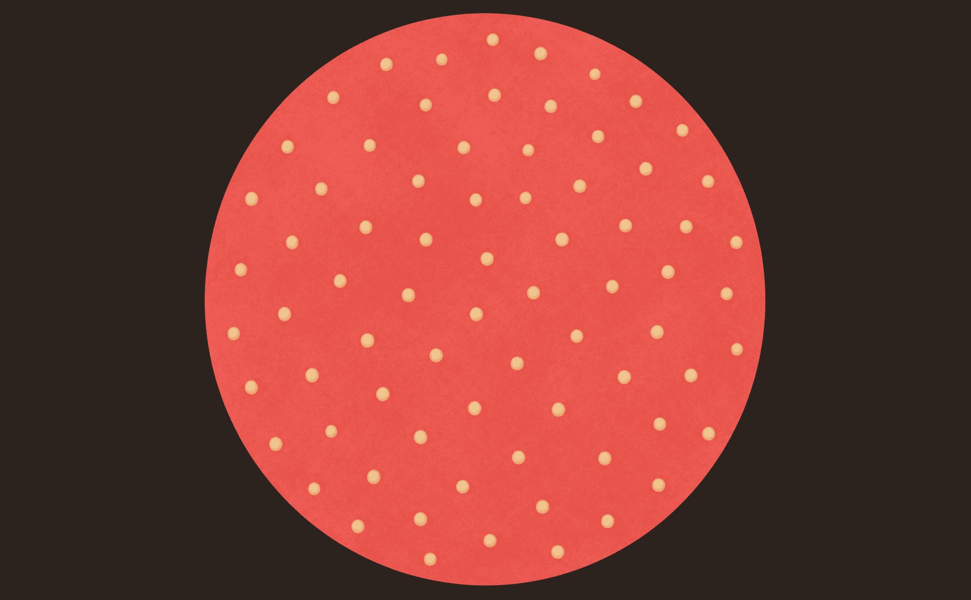
Strawberry pattern: pink-red background studded with solid white-yellow follicular plugs — the dotted surface resembling a strawberry
Associated with: Actinic keratosis (characteristic feature). See Non-Melanoma Skin Cancer.
Structureless areas
Plain language: Areas of colour without any distinct pattern or structure.
What you see: Zones of homogeneous colour — brown, grey, pink, white, or blue — without network, globules, dots, or other discrete features. The significance depends entirely on the colour and clinical context.
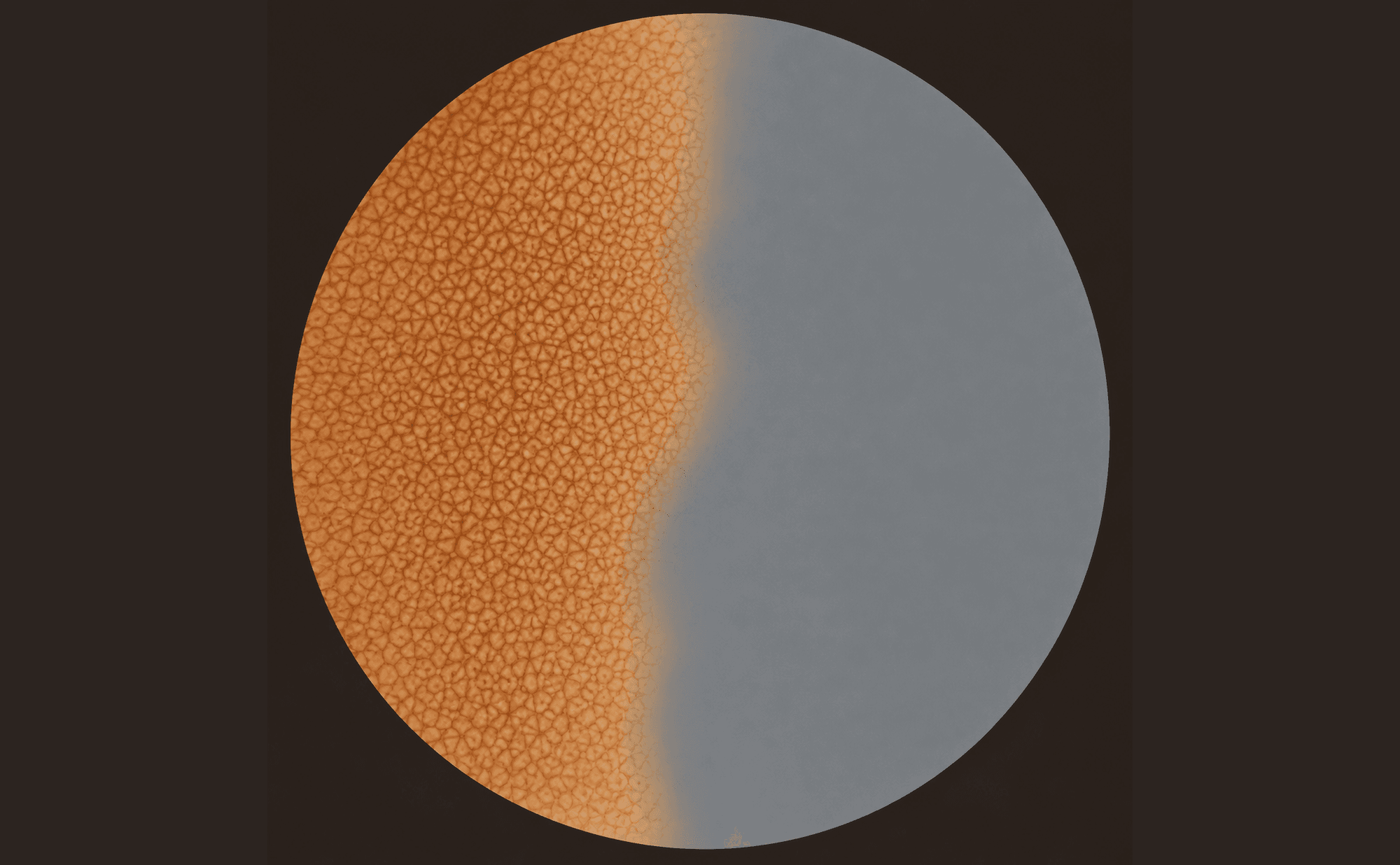
Structureless areas: a zone of flat, homogeneous colour with no network, dots, or lines — shown here against a structured zone for contrast
Associated with: Various — interpretation depends on colour and lesion context. See Homogeneous pattern (above).
T
Targetoid hair follicles
Plain language: Hair follicles with concentric colour rings — darker centre, lighter surround.
What you see: Follicular openings with concentric colour variation creating a target-like or bullseye appearance around each follicle.
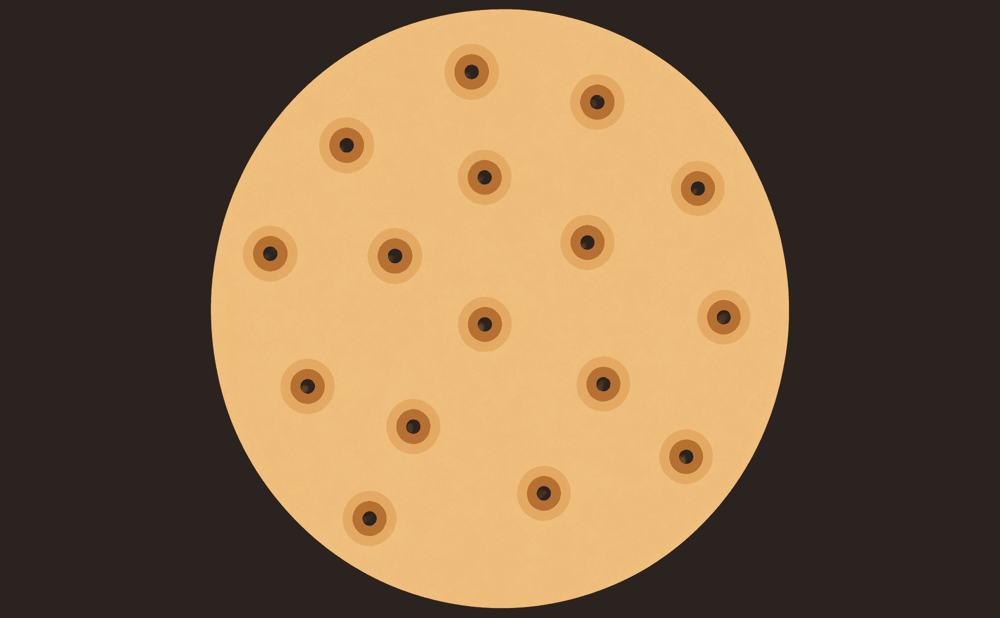
Targetoid hair follicles: concentric colour rings around each follicular opening — a target/bullseye appearance
Associated with: Lentigo maligna, Bowen's disease. See Malignant Patterns and Non-Melanoma Skin Cancer.
U
Ulceration
Plain language: Loss of the skin surface with crusting or bleeding.
What you see: Disruption of the epidermis with erosion, crusting, or exposed underlying tissue. May have dried blood. Indicates the lesion has outgrown or destroyed the overlying skin. One of the seven TADA high-risk features.
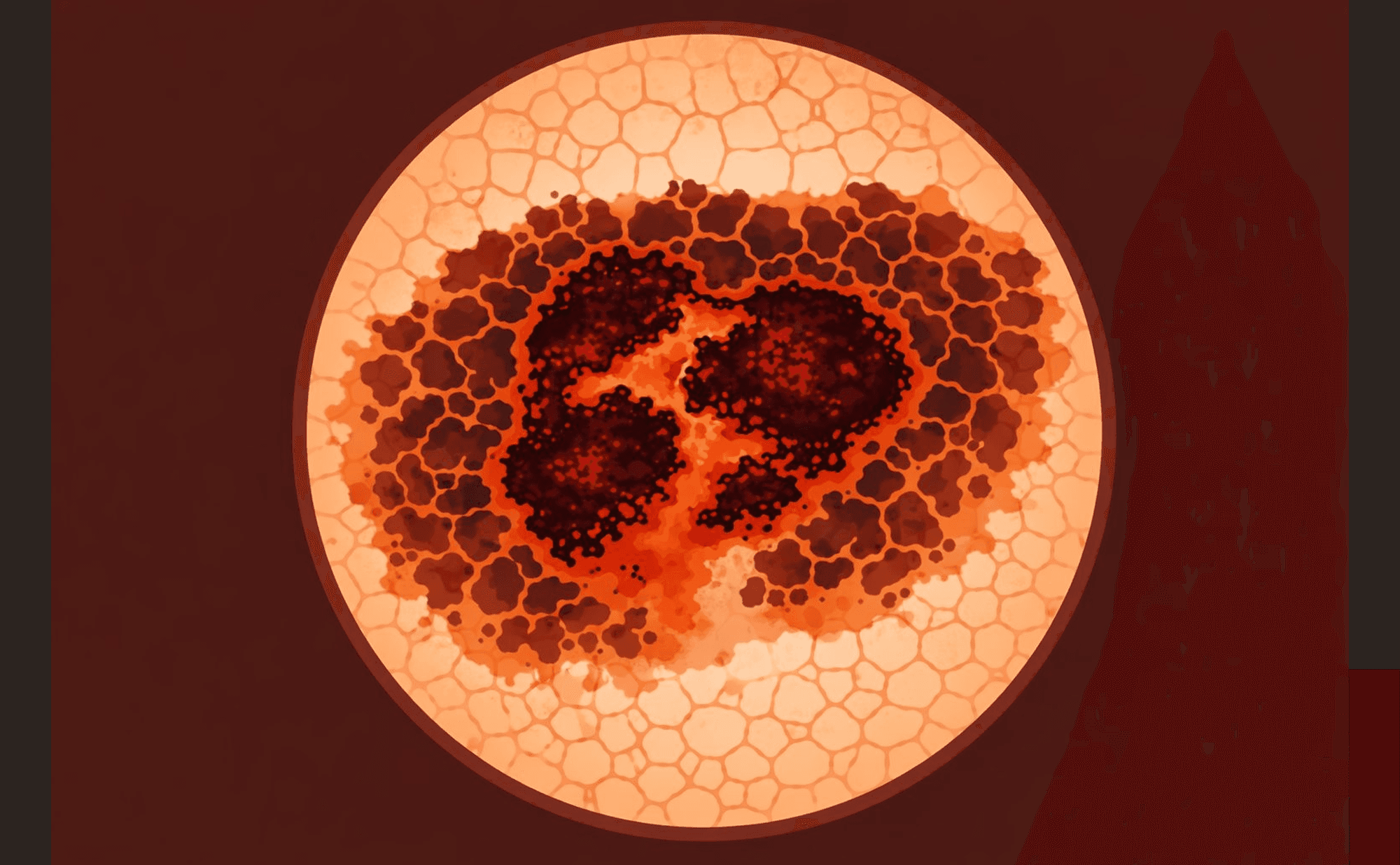
Ulceration: epidermal disruption with crusting and erosion — one of the seven TADA high-risk features
Associated with: Melanoma (advanced), BCC, SCC, other malignancies. See Malignant Patterns and Non-Melanoma Skin Cancer.
W
White network
Plain language: Grid pattern of white lines — different from negative network.
What you see: A network pattern where the lines are white rather than brown. Often seen centrally in dermatofibroma, where it reflects the underlying fibrosis pushing the epidermis upward.
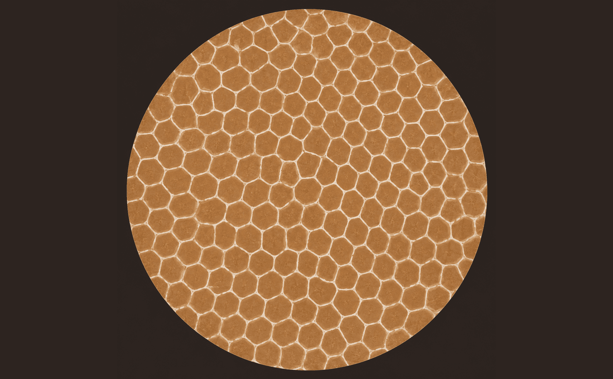
White network: a regular grid of white lines rather than brown — often seen centrally in dermatofibroma
Associated with: Dermatofibroma. See Benign Patterns.
Quick reference by lesion
Seborrhoeic keratosis
Comedo-like openings · Milia-like cysts · Fissures and ridges · Hairpin vessels · Sharp demarcation
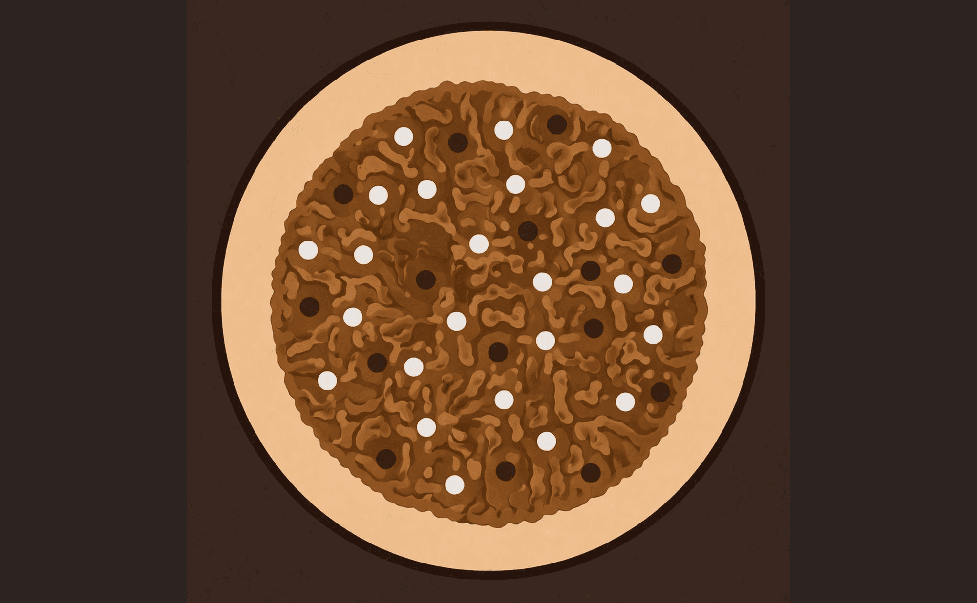
Classic seborrhoeic keratosis: milia-like cysts, comedo-like openings, cerebriform fissures and ridges, and sharp demarcation
Cherry angioma
Red lacunae · Pale septa · Lacunar pattern · Sharp demarcation
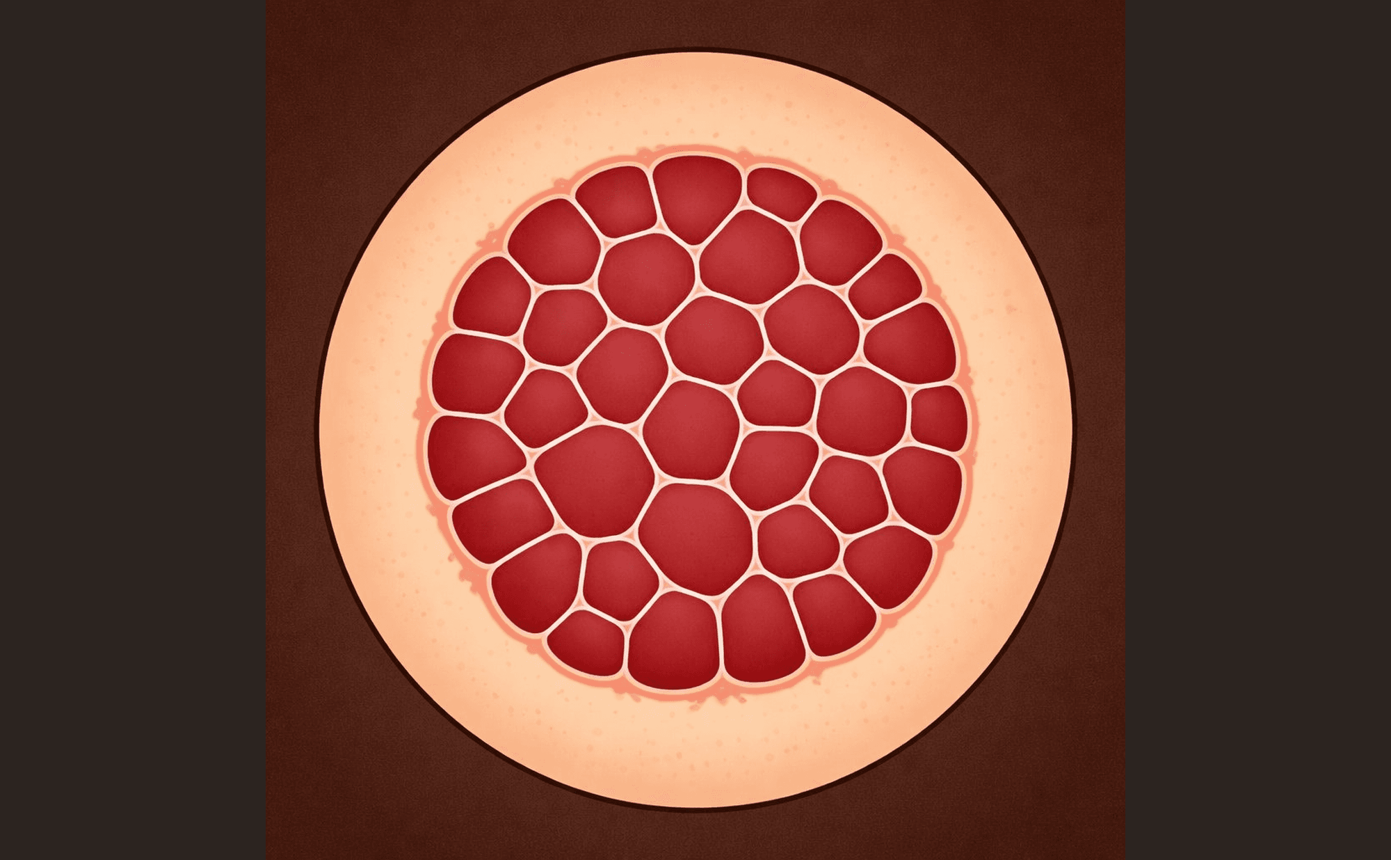
Classic cherry angioma: red-purple lacunae separated by pale septa — the stained-glass-window pattern
Dermatofibroma
Central white patch · Peripheral pigment network · White network (variant) · Dimple sign (clinical)
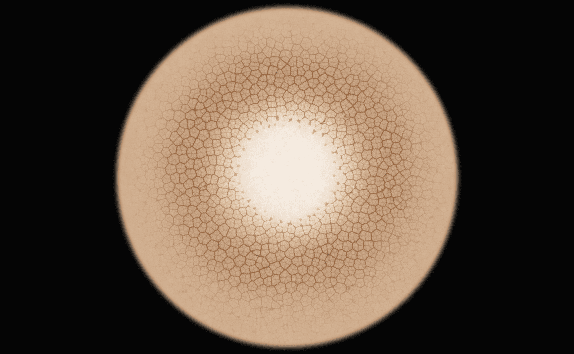
Classic dermatofibroma: central white scar-like zone with peripheral pigment network — the bullseye pattern
Melanoma (superficial spreading)
Atypical network · Irregular dots and globules · Asymmetry · Blue-white veil · Regression structures · Multiple colours
Nodular melanoma
Blue-black colour · Polymorphous vessels · Ulceration · Shiny white structures
Basal cell carcinoma
Arborising vessels · Shiny white structures · Blue-grey ovoid nests · Leaf-like areas · Spoke-wheel structures · Ulceration
SCC / Bowen's disease
Glomerular vessels · Keratin and scale · Hairpin vessels · White circles · Ulceration (invasive)
Actinic keratosis
Strawberry pattern · Rosettes · White follicular openings · Surface scale